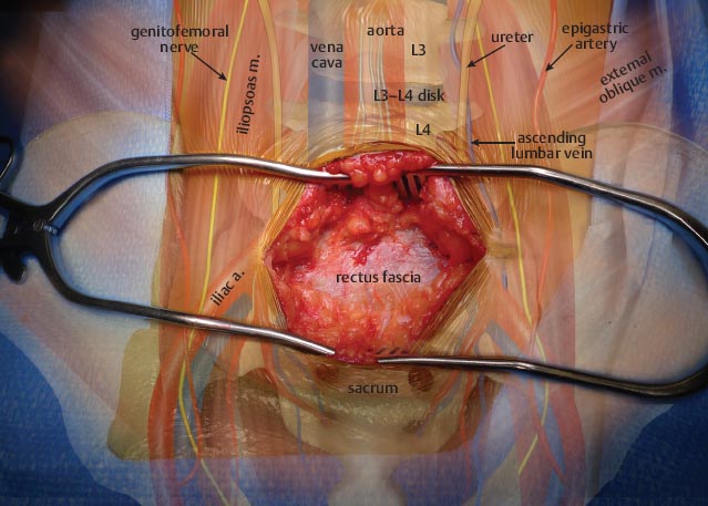
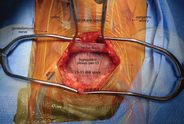
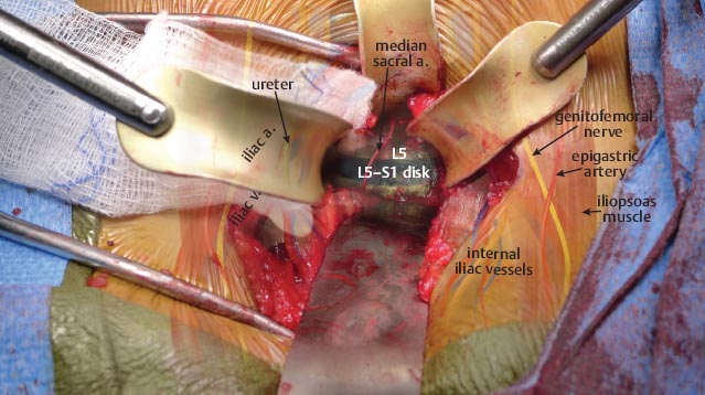
 Exposure and Approach
• A lateral fluoroscopic image should be obtained before incision to localize the surgical level. At the L5-S1 level, the great vessels (aorta/vena cava) have bifurcated. At the L4-L5 disk space level, the great vessels are retracted to the right. At L4-L5, the ascending lumbar vein may need to be ligated to mobilize the vessels. Once the incision has been made, the fascia of the musculus rectus abdominis is incised. The fascial incision can made either horizontally (in line with the skin incision) or vertically, depending upon the surgeon’s preference.

• Rectus fascia identified.

• The muscle belly of the rectus is mobilized. Some surgeons argue that mobilizing the lateral edge of the rectus results in denervation of the muscle, while others state that medial mobilization increases the likelihood for abdominal hernias. Posterior to the rectus is the rectus sheath, which is incised carefully, exposing the retroperitoneum.

Stay updated, free articles. Join our Telegram channel

Full access? Get Clinical Tree


