Fig. 18.1
Atypical pituitary adenoma. (a) Sagittal T1-weighted post-gadolinium image. (b) Coronal T1-weighted post-gadolinium image. A large, heterogeneously enhancing sellar mass demonstrates suprasellar extension and encasement of the right cavernous internal carotid artery, as well as erosion of the sellar floor. Superiorly, the mass has elevated the floor of the third ventricle, and the optic chiasm is likely elevated and compressed. Posteriorly, the mass effaces the prepontine cistern and mildly impresses on the pons. The sagittal image incidentally shows a small pineal cyst
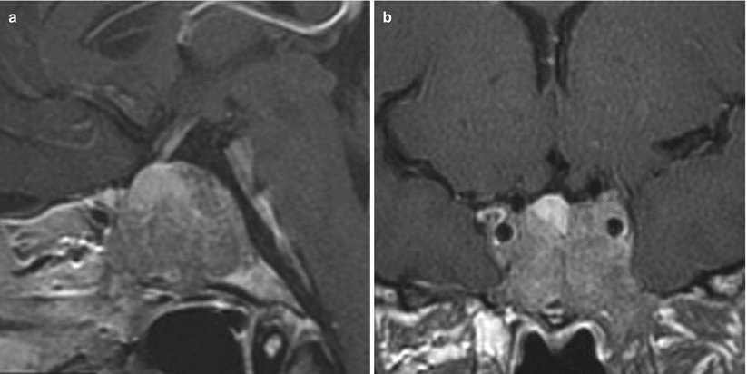
Fig. 18.2
Atypical pituitary adenoma. (a) Sagittal T1-weighted post-gadolinium image. (b) Coronal T1-weighted post-gadolinium image. A large, heterogeneously enhancing sellar mass encases the cavernous internal carotid arteries bilaterally, and there is erosion of the sellar floor. The normal pituitary glandular tissue is seen along the superior margin of the mass
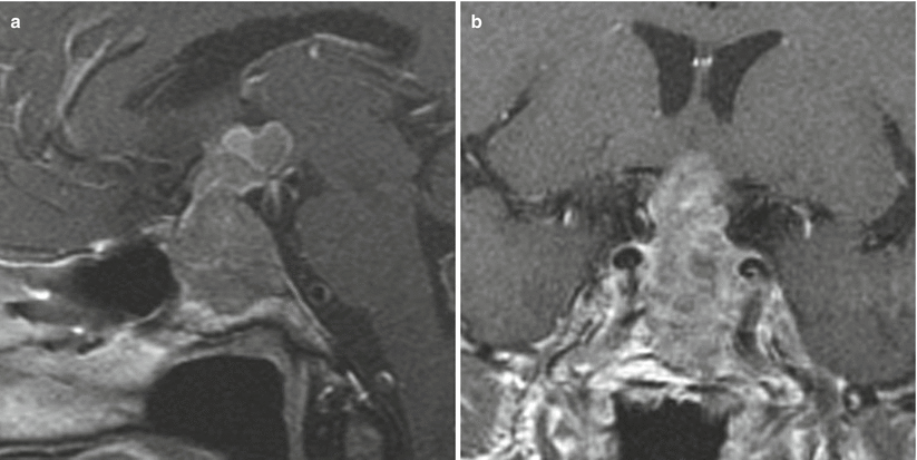
Fig. 18.3
Atypical pituitary adenoma. (a) Sagittal T1-weighted post-gadolinium image. (a) Coronal T1-weighted post-gadolinium image. There is a large, heterogeneously enhancing sellar mass with suprasellar extension. The mass abuts the cavernous internal carotid arteries without definite encasement. There is erosion of the sellar floor. Superiorly, the mass has elevated the floor of the third ventricle, and the optic chiasm is likely elevated and compressed
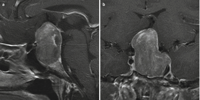
Fig. 18.4
Atypical pituitary adenoma. (a) Sagittal T1-weighted post-gadolinium image. (b) Coronal T1-weighted post-gadolinium image. A large, heterogeneously enhancing sellar/suprasellar mass abuts the cavernous internal carotid arteries, with possible invasion of the left cavernous sinus on the left. There is erosion of the sellar floor. Superiorly, the mass has elevated the lamina terminalis, and the optic chiasm is likely elevated and compressed
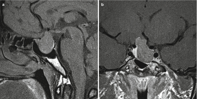
Fig. 18.5
Atypical pituitary adenoma. (a) Sagittal T1-weighted post-gadolinium image. (b) Coronal T1-weighted post-gadolinium image. There is a heterogeneously enhancing sellar mass with suprasellar extension. The mass abuts the left cavernous internal carotid artery without definite invasion of the left cavernous sinus. There is erosion of the left sellar floor. Superiorly, the mass abuts the right hypothalamus. The normal pituitary glandular tissue is seen along the right aspect of the sella
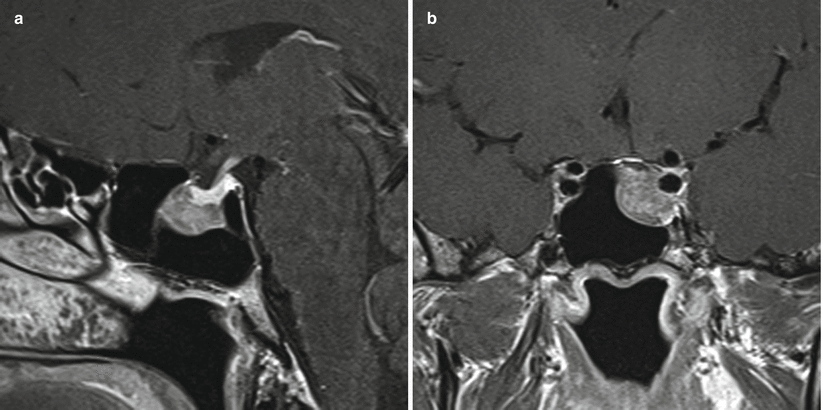
Fig. 18.6
Atypical pituitary adenoma. (a) Sagittal T1-weighted post-gadolinium image. (b) Coronal T1-weighted post-gadolinium image. An enhancing sellar mass in the left aspect of the sella abuts the left cavernous internal carotid artery without definite invasion of the left cavernous sinus. There is erosion of the left sellar floor. The normal pituitary glandular tissue is seen along the superior aspect of the mass
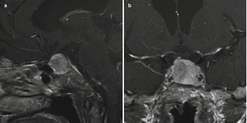
Fig. 18.7


Atypical pituitary adenoma. (a) Sagittal T1-weighted post-gadolinium image. (b) Coronal T1-weighted post-gadolinium image. There is an enhancing sellar mass in the sella, abutting the cavernous internal carotid arteries bilaterally without definite invasion of the cavernous sinuses. There is erosion of the left sellar floor. The normal pituitary glandular tissue is seen along the superior aspect of the mass
Related posts:


Stay updated, free articles. Join our Telegram channel
Full access? Get Clinical Tree
