Fig. 51.1
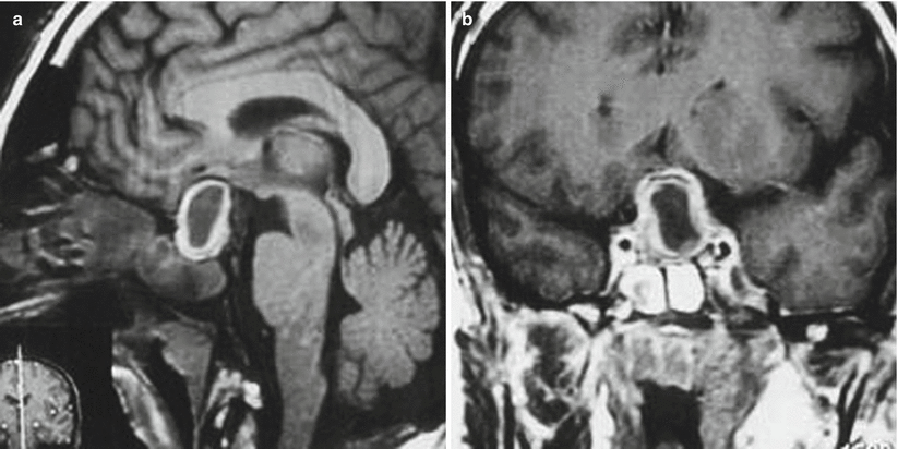
Bacterial sellar abscess. Preoperative MR images of a patient with a history of remote prior transsphenoidal surgery for a pituitary tumor and a bacterial sellar abscess: Sagittal (a) and coronal (b) T1-weighted images demonstrate an isointense lesion in the pituitary fossa, with ring enhancement after contrast injection (Adapted from Ciapetta et al. [20] with permission)

Fig. 51.2
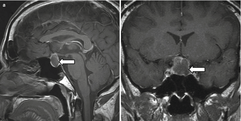
Spontaneous pituitary abscess. Preoperative (postcontrast) MR images in a patient with a spontaneous pituitary abscess. (a) Sagittal section shows a sellar cystic mass with suprasellar extension (arrow). The lesion is seen pushing the optic chiasm upward. The pituitary stalk is intact. (b) Coronal section shows the pituitary sellar mass with suprasellar extension (arrow). The lesion is seen pushing the optic chiasm upward (Adapted from Dalan and Leow [14], with permission)

Fig. 51.3
Spontaneous abscess. Preoperative post-gadolinium T1-weighted MRI scans in a patient with a spontaneous aspergillosis abscess. A sellar and suprasellar mass with peripheral contrast enhancement is seen on sagittal (a) and coronal (b) sections. Marked compression of the optic chiasm and thickened sphenoid sinus mucosa (suggesting sinusitis) can also be seen (Adapted with permission from Iplikcioglu AC et al., Acta Neurochir (Wien) (2004) 146:521–524)
51.3 Histopathology
A wide variety of bacterial and fungal organisms have been implicated in the formation of sellar abscesses.
Histopathology may demonstrate necrosis and abscess wall, with infiltration by polymorphonuclear leukocytes or macrophages [2].
In most cases (over 80 %), organisms are not isolated on cultures [7].
Polymerase chain reaction (PCR) techniques may be useful in establishing a definitive diagnosis.
51.4 Clinical and Surgical Management
The diagnosis of sellar abscess is often missed prior to surgery [7].
Transsphenoidal surgical drainage and marsupialization of the abscess wall is the standard management [7, 20].
In some cases, conservative management with antibiotics may be attempted, although there is little evidence to support this strategy [21].
Intraoperative cultures for aerobic, anaerobic, fungal, and acid-fast bacilli should be sent.
Sellar floor reconstruction is not recommended unless a CSF leak is present.
For bacterial abscesses, intravenous antibiotics are typically required for at least 6 weeks after drainage.
Tubercular or fungal abscess should be treated with the appropriate antibiotic regimens.
Symptoms from mass effect generally improve following drainage, whereas hypopituitarism typically fails to do so [12, 22].
References
1.
2.
Dutta P, Bhansali A, Singh P, Kotwal N, Pathak A, Kumar Y. Pituitary abscess: report of four cases and review of literature. Pituitary. 2006;9:267–73.CrossRefPubMed
Stay updated, free articles. Join our Telegram channel

Full access? Get Clinical Tree


