Basic Cellular Neurophysiology


Basic Cellular Neurophysiology


Stephen W. Jones


ION CHANNELS AND INTRINSIC MEMBRANE PROPERTIES


Basis of Electrical Activity

The activity of ion channels is fundamental to signaling in the nervous system. Synaptic potentials and action potentials, the basic electrical signals, result directly from the movement of ions through channels. In past decades, patch clamp recording and molecular biology have identified a variety of ion channel types and have begun to clarify the mechanisms underlying channel activity.

The electrical activity of a biologic membrane can be represented as an equivalent circuit consisting of a resistor, a capacitor, and a battery (Fig. 5.1A). This corresponds physically to ion channels, the lipid bilayer, and ion gradients (Fig. 5.1B). The properties of the bilayer and the ion gradients are essentially constant, with the important exception of the Ca2+ gradient. In contrast, ion channels vary dramatically among cell types and can open or close in response to physiologic stimuli. The intrinsic membrane properties of a cell and its moment-to-moment electrical activity depend primarily on the types and numbers of ion channels that the cell expresses.

How do the channels, bilayer, and ion gradients work together to produce a membrane potential? A pure lipid bilayer is an excellent insulator, because its hydrophobic interior renders it essentially impermeable to ions. Movement of ions across the membrane (electrically, a current) occurs primarily through ion channels. Because an open ion channel catalyzes the flux of ions down a concentration gradient, it is more intuitive to think in terms of conductance rather than resistance: an increase in the number of open channels produces a higher electrical conductance. The energy source for ion flow through a channel is an ion gradient, resulting from the activity of pumps and carriers, such as the Na+ ,K+ adenosine triphosphatase (ATPase). Ion flow changes the charge distribution across the membrane. Because like charges repel each other, the presence of net charge in bulk solution is an unstable situation (i.e., principle of electroneutrality). The lipid bilayer is extremely thin (7 nm), allowing strong attractive forces between positively and negatively charged ions on opposite sides of the membrane. This allows the bilayer to act as a capacitor to store charge. The separation of charge across the membrane is the source of the membrane potential (VM): VM = Q/C, where Q is the amount of charge, and C is the capacitance of the membrane. Biologic membranes have an essentially constant capacitance, approximately 1 μF/cm2.

The membrane potential depends primarily on the selectivity of the ion channels that are open at a given time. Ion channels can be highly selective for particular ions. For example, many K+ channels are more than 100-fold more selective for K+ than for Na+. For a cell with only K+ channels open (i.e., an approximation of a real neuron at its resting potential), a chemical or diffusional force allows K+ to diffuse down the concentration gradient and out of the cell. As K+ ions flow, however, the outside (o) of the cell becomes electrically positive with respect to the inside (i). Convention defines the voltage outside a cell as zero, so the membrane potential resulting from outward movement of K+ ions is said to be negative. That voltage produces an electrical force that prevents further efflux of K+ ions, as the excess of positive charge outside the cell repels the positively charged K+. If no other ions can cross the membrane, a state of equilibrium is rapidly reached in which the outward diffusional force equals the inward electrical force. That would be a true thermodynamic equilibrium, stable indefinitely without input of metabolic energy. There would be no net flux of K+ through an open K+ channel. The voltage across the membrane would be the equilibrium potential for K+ (EK), as given by the Nernst
equation for a battery: at 37°C, EK = 61.5 log([K+]o/[K+]i). EK is near −90 mV for a physiologic K+ gradient.


Figure 5.1 Electrical (A) and physical (B) views of a K+-selective membrane.

By the same argument, if only Na+-selective channels are open, the membrane potential would be ENa, or about +60 mV. That is a first approximation to a neuron at the peak of the action potential.

In reality, the situation is more complex, because multiple channel types are open simultaneously. There is no generally valid way to calculate the membrane potential in that case, although two equations are commonly used. The more familiar is the Goldman equation:

VM = 61.5 log {(PK [K+]o + PNa [Na+]o) / (PK [K + ]i + PNa [Na+]i)}

in which PK and PNa are the permeabilities of the membrane to K+ and Na+ (permeabilities are related to conductances). The derivation of the Goldman equation uses some assumptions that are not realistic for ion channels, particularly that ions move independently of one another, but it is a useful approximation and often serves as an empirical definition of ion permeability.

In another approach, Ohm’s law is used to define the conductance for each ion: for K+, IK = GK (VM − EK). The “driving force,” the difference between the membrane potential and the equilibrium potential, is used rather than the absolute voltage, because the current through an open K+ channel goes to zero at the equilibrium potential. If the membrane potential is constant, there must be no net ionic current: IK + INa = 0 (considering only K+ and Na+). That leads to the “parallel batteries” equation:

VM = G′K EK + G′Na ENa

The equation uses normalized conductances G′K = GK/(GK + GNa) and G′Na = GNa/(GK + GNa). Qualitatively, the Goldman equation and the parallel batteries equation say that the membrane potential is a compromise among the equilibrium potentials for the individual ions. The higher the conductance (or permeability) for an ion, the closer the membrane potential is to that ion’s equilibrium potential. This allows a better approximation to the situation at rest: the membrane potential is much closer to EK than to ENa because many more K+ channels are open than Na+ channels. The converse is true at the peak of the action potential. For completeness, there is also a delay between channel opening and a change in voltage, because time is required for ion flow to change the net distribution of charge across the membrane (i.e., to charge the membrane capacitance).


General Properties of Ion Channels

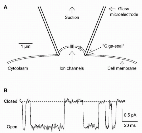
Until 25 years ago, ion channels were considered hypothetical entities. The most direct demonstration of discrete ion channels came from patch clamp recording (Fig. 5.2A). When a glass electrode with an opening of about 1 μm is pressed onto a cell surface, gentle suction often forms a seal between the glass and the cell membrane that is tight electrically, chemically, and physically. The resistance of the seal is typically about 10 GΩ (1010 Ω). That extremely low rate of ion leakage under the seal implies molecular tightness. The high resistance of the seal gives very low levels of current noise, so that the tiny currents resulting from activity of individual ion channels in the “patch” of membrane directly under the electrode can be resolved. This was one of the first scientific techniques by which the behavior of a single molecule in real time could be observed.

Two types of information can be obtained from single-channel records (Fig. 5.2B): gating (i.e., when is a channel open?) and permeation (i.e., how does current flow through an open ion channel?). A channel’s permeation properties determine its ion selectivity. The absolute amplitude of single-channel currents is also instructive. A typical 2-pA
current may seem inconsequential, but it translates into a flux of 107 ions per second through the channel pore. That simple calculation remains one of the strongest pieces of evidence that an ion channel is a channel, because that transport rate is orders of magnitude higher than the turnover number of even the fastest enzymes and carriers, and the only known mechanism that could produce such a rate is diffusion through a pore, with movement at a rate nearly as fast as if the ions were in bulk water. The combination of high flux and high selectivity among chemically similar ions remains an interesting puzzle.


Figure 5.2 The patch clamp technique. A: A patch electrode is sealed onto a cell. This cell-attached configuration records the activity of individual ion channels located in the patch of membrane under the electrode. B: Example of ion channel gating for an L-type calcium channel from a smooth-muscle cell. (Courtesy of Dr. Carlos A. Obejero-Paz, Case Western Reserve University, Cleveland, OH.)


Figure 5.3 Two possible mechanisms of channel gating. A: Movement of a “gate” can cause the channel pore to be open or blocked at one end, as is thought to occur for rapid inactivation of A-type potassium channels. B: Channel opening also may result from a concerted conformational change involving the entire protein. This diagram shows net outward movement of positive charges within the channel protein as the channel opens, which would produce a voltage-dependent channel that opens on depolarization.

Channel gating is crucial, because the opening and closing of ion channels in response to physiologic stimuli is the fundamental mechanism underlying signals such as synaptic potentials and action potentials. What is gating? Some general conclusions can be drawn from records of single channels (
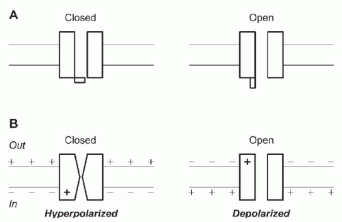
Fig. 5.2B). Channels switch between open and closed states, apparently at random. Under fixed recording conditions, the current observed when a channel is open generally is constant, implying a constant rate of ion movement through the pore. The current through a closed channel is almost undetectable. The transition between the open and closed states is too fast to measure, implying a completed conformational change in a few microseconds or less. Two extreme physical pictures can be used to illustrate gating: literal movement of a gate, a part of the channel protein that can occlude the pore or move out of the way (Fig. 5.3A), or a global conformational change in the protein (Fig. 5.3B). Both mechanisms probably occur.

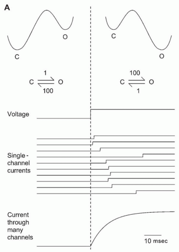
The observed random opening and closing of channels are precisely what would be expected when a chemical reaction is viewed one molecule at a time (Fig. 5.4). The simplest possible reaction scheme, the closed-open (C-O) model, with first-order transitions linking closed and open states, predicts that the channel will be open a particular fraction of the time, depending on the equilibrium constant for the reaction. The average amount of time that the channel stays in the closed state depends on the rate constant for the C → O reaction. The closed times are highly variable, because the channel must “wait” until thermal energy “pushes” it over the barrier linking the two states. Random does not mean causal, however. Although the duration of any individual closing event is unpredictable, the average behavior of the channel is determined by the rate constants in the kinetic scheme. Real ion channels can exist in considerably more than two states, but the simple two-state C-O model is the starting point for discussion of channel kinetics.


Figure 5.4 The simplest model for channel gating. Two conformational states of the protein (closed [C] and open [O]) spontaneously interconvert according to first-order rate constants k1 and k−1. The right-hand diagram shows the energy levels of the closed and open states, which determine the equilibrium, and the height of the energy barrier, which determines the rates.

Although useful information can be obtained by recording channel behavior under fixed conditions, the response of channels to changes in conditions allows signaling in the nervous system. Two main factors affect the rates of channel opening and closing: binding of ligands (e.g., neurotransmitters) and voltage. Ligand-gated channels and voltage-dependent channels are functionally and molecularly distinct. The simplest scheme for a ligand-gated channel is a bimolecular binding reaction (Fig. 5.5), in which binding and channel opening are considered to be a single step. This model makes firm predictions: the channel never opens in the absence of ligand, and the channel’s opening rate increases linearly with the ligand concentration. The channel’s mean closed time (for this scheme, the reciprocal of the opening rate) decreases with concentration, and its mean open time does not depend on concentration. As the concentration of ligand increases, the time that the channel has to wait before a ligand binds decreases, enhancing the probability that the channel is open. At very high concentrations, channel closings are extremely brief. These predictions can be tested experimentally. If the tests fail, more states are added to the model, with the ultimate goal being to gain physical insight into the mechanism of channel activation.

Why ion channel gating would depend on voltage is less obvious. Typical membrane potentials are a few tens of
millivolts, but the voltage drop occurs across the extremely thin lipid bilayer. A 70-mV voltage for a 7-nm-thick membrane produces an electrical field of 107 V/m, nearly enough to electrolytically break down even as good a capacitor as a lipid bilayer. The electrical forces on any charged groups in a membrane protein are extremely strong if those charges are in the membrane’s electrical field. That suggests a simple mechanism for voltage dependence. If the closed and open states of a channel differ in their distribution of charge across the membrane (Fig. 5.3B), the equilibrium between the two states will depend on voltage. Only a small number of charges have to move to change the open probability of a channel from near 0 to near 1 over the physiologic range of membrane potentials.


Figure 5.5 Simulation of a ligand-gated channel exhibiting two-state (C-O) gating. The channel-opening step is a bimolecular reaction between the ligand (e.g., neurotransmitter) and the channel. The probability that the channel is open increases with concentration, because the pseudo-first-order rate constant for channel opening (k1) is proportional to the ligand concentration.

When the voltage changes, voltage-dependent channels do not instantly change from one state to another. Consider the two-state C-O channel (Fig. 5.6A). Suppose that at a negative voltage the rate constants are such that the channel spends 99% of its time closed. For the numbers given, on average, the channel would remain closed for 1 second, and then open for 10 milliseconds before closing again. Suppose that at a more positive voltage the rate constant for channel opening increases and that for closing decreases by 100-fold (for a voltage-dependent channel, both opening and closing rates usually change). The channel will spend 99% of its time open at equilibrium. If the voltage is changed instantly from negative to positive, the rate constants will change instantly, but the channel will require some time to open, as determined by the “new” rate constant. Figure 5.6A illustrates a simulated voltage clamp experiment in which a patch containing a single channel is repeatedly depolarized. At the negative voltage, the channel is nearly always closed at equilibrium. On depolarization, the channel waits a random time (mean, 10 milliseconds) before accumulating enough energy to navigate the barrier to the open state. At equilibrium, the channel is nearly always open at the depolarized voltage. If the experiment is repeated many times, or if an analogous voltage clamp experiment is done on all of the cell’s channels, an exponential relaxation from a p(open) of 0.01 to 0.99 will result, with a time constant of 10 milliseconds. This is a first approximation to the behavior of a voltage-dependent K + channel of the type involved in the action potential. The rates of channel opening and closing depend on the
height of the energy barriers at each voltage. The absolute rates vary widely among channel types.


Figure 5.6 Simulation of channel gating for simplified K+ and Na+ channels. A: Response of a two-state K+ channel to depolarization. The rate constants favor the closed state at negative voltages and the open state at depolarized voltages. The latency to first opening of a channel is the reciprocal of the opening rate constant (here, 10 milliseconds). The traces labeled single-channel currents show 10 simulated responses of a channel to depolarization; the current through many channels shows the predictable macroscopic behavior of a cell containing many thousands of channels. B: Response of a three-state Na+ channel to depolarization. All the voltage dependence of the channel is assumed to be in the opening and closing rates. For such a model, the apparent voltage dependence of inactivation comes from kinetic coupling between the activation and inactivation steps. This adequately describes the channel’s response to depolarization but not the recovery from inactivation. For these parameters, the channels usually open only once during a brief depolarization and occasionally fail to open at all, because there is a small probability that the channel will be inactivated (I) at the hyperpolarized holding potential.


Figure 5.6 (Continued)

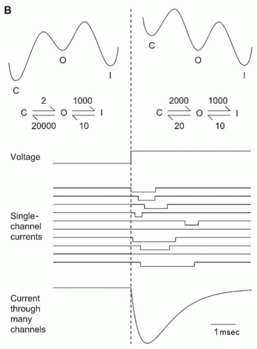
The gating of a voltage-dependent Na+ channel differs in two crucial ways from that of a K+ channel. First, although it also opens on depolarization, the rate constant for opening is approximately 10-fold faster than for the K+ channel. Second, during a maintained depolarization, the Na+ channel does not remain open. Because this cannot be explained by the C-O model, at minimum, a third inactivated (I) state must be added to the kinetic scheme (Fig. 5.6B). That state is also closed in the sense that no ions go through the channel, but the closed and inactivated states are distinct conformational states of the channel protein.

The next step is to explain how channel opening and closing result in electrical signals. A discussion of the mechanism of the action potential follows.


Action Potential

When a cell with Na+ and K+ channels is depolarized, both types of channels open, but Na+ channels open more rapidly. The influx of Na+, net inward movement of positive charge, makes the interior of the cell more positive than before; it depolarizes the cell. The depolarization opens more Na+ channels, giving more Na+ entry and causing more depolarization (Fig. 5.7). This positive feedback cycle initiates the action potential and is responsible for its all-or-none character. If the cell has sufficient Na+ channels, the current generated greatly exceeds the stimulus necessary to trigger the action potential. Once initiated, the action potential is independent of the stimulus.


Figure 5.7 Positive and negative feedback resulting from opening of voltage-dependent channels. If enough Na+ channels open to produce net inward current (i.e., more inward Na+ current than outward K+ current), the resulting depolarization will open even more Na+ channels, and an action potential will result. In contrast, K+ channel opening hyperpolarizes a cell.

Termination of the action potential, called repolarization, involves two simultaneous processes. Na+ channels inactivate, and K+ channels open. Opening of K+ channels provides negative feedback (Fig. 5.7); efflux of K+ from the cell makes the inside more negative, which tends to close K+ channels. The negative feedback contributes to repolarization.

The resting K+ conductance of the membrane stabilizes the resting potential. Very small depolarizations do not trigger an action potential. They do not open a large number of Na+ channels but increase the driving force on K+ (because a depolarized membrane is further from EK) and decrease the driving force on Na+. The net effect of a small depolarization is to increase K+ efflux, which leads to hyperpolarization. A sharp boundary, the threshold, exists between small depolarizations that produce only a subthreshold response and slightly larger depolarizations that trigger a full-sized action potential. Threshold is the point at which inward Na+ current exceeds outward K+ current; the net inward current produces an active depolarization, which initiates further Na+ channel opening and the rest of the positive feedback cycle.

The kinetic differences between voltage-dependent Na+ and K+ channels are crucial to the action potential. During repolarization, K+ channel opening and Na+ channel inactivation cooperate. Because K+ channels also close slowly, the K+ conductance of the membrane immediately after repolarization is higher than at rest. The membrane potential is very close to EK, producing an afterhyperpolarization (AHP).


Hodgkin-Huxley Experiments and Model

Our detailed understanding of the mechanism of the action potential is based on the classic experiments of Hodgkin
and Huxley on the giant axon of the squid (1). Current views of the mechanisms of channel gating owe much to the kinetic models they proposed in 1952. That is remarkable, given that the structure of biologic membranes was then poorly understood. Although lipids and proteins were known to be involved, the idea of transmembrane proteins was yet to come. Nevertheless, their descriptions of “permeability changes” can be translated directly into the opening and closing of ion channels. As discussed later, contemporary views of channel gating differ from these original models, but the similarities are impressive.

The Hodgkin-Huxley experimental protocols are the foundation for analysis of ion channel gating to this day. Their basic experiment was to give step changes in voltage (Fig. 5.6). That makes perfect sense now: if a channel is voltage dependent, the first step toward understanding it is to control the relevant variable, the membrane potential. They first defined two kinetically distinct and independent pathways for ion movement, which we now interpret as Na+ and K+ channels. Ion substitutions played a major role in their separation of Na+ and K+ currents. For example, Hodgkin and Huxley demonstrated that the Na+ current reversed from an inward flow of Na+ to an outward current when the axon was depolarized beyond the expected ENa. That “reversal potential” shifted, as predicted by the Nernst equation, when ENa was changed by replacing some of the extracellular Na+ with an impermeant cation. The reversal potential remains the primary criterion for determining ion channel selectivity. Hodgkin and Huxley also characterized the kinetics of channel opening and closing over the physiologic voltage range. They discovered that Na+ channels inactivate and described the voltage dependence of that process.

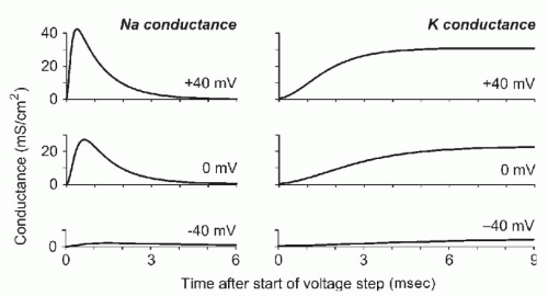
Figure 5.8 summarizes much of their experimental data. Because conductance is roughly proportional to the probability that a channel is open, their data on Na+ and K+ conductance as a function of voltage and time reflect the kinetics of channel gating. In some ways, the K+ and Na+ conductances behaved as expected from the previously discussed C-O and C-O-I models (Fig. 5.6), but there were complications. In particular, the time course of activation of the currents was not exponential but showed a sigmoid delay, necessitating more complex kinetic models. No general strategy was available to translate data into a model, and computational limitations encouraged keeping the models as simple as possible.


Figure 5.8 The time course for opening of Na+ and K+ channels, as calculated from the Hodgkin-Huxley model (1). The conductance is proportional to the number of channels open and is expressed (per cm2 of membrane) in units of Siemens (S), or reciprocal ohms (Ω−1). Notice that stronger depolarization activates more Na+ and K+ channels and that the channels open more rapidly.

Hodgkin and Huxley modeled Na+ and K+ channels as containing four independent gates (1). Each gate behaves kinetically like a C-O channel, but the channel is assumed to be open if and only if all four gates are open simultaneously. The K+ channel model is simpler because it is assumed that the four gates are identical. The assumption that the gates are independent simplifies matters, because independent probabilities multiply. If the probability that one gate is open is n, then the probability that a channel is open is n4. The requirement for all four gates to open produces the sigmoid delay in the time course of activation of the K+ channel (Fig. 5.8).

To explain Na+ inactivation, Hodgkin and Huxley assumed that one of the four Na+ channel gates is an inactivation gate, with a reverse voltage dependence; it is open at negative voltages and closes at depolarized voltages. The three activation gates open on depolarization, more rapidly than the gates controlling a K+ channel. In their equations, m is the probability that a Na+ activation gate is open and h the probability that the inactivation gate is open, so that the probability that a Na+ channel is open is m3h. The Hodgkin-Huxley model included empirical equations for the voltage dependence of the rate constants for each gate’s opening and closing. That allowed calculation of m, h, and n (and from them, the probabilities that Na+ and K+ channels are open) as functions of voltage and time. Their model reproduced the experimental data obtained under voltage clamp conditions and generated an action potential when the membrane voltage was not controlled. Like a real action potential, the computed action potential had a threshold, the ability to propagate, and a refractory period.


CONTEMPORARY WORK ON VOLTAGE-DEPENDENT ION CHANNELS

Since 1952, we have relearned something that Hodgkin and Huxley recognized, that different neurons can have dramatically different patterns of electrical excitability. One striking example is in leech sensory neurons (2). Three types of cells—called T, P, and N—can be identified from animal to animal on the basis of their anatomy and receptive field properties. They can also be distinguished by the shape of their action potentials. Their responses to a maintained depolarization differ systematically; for example, the N (nociceptive) cells have higher thresholds and less of a tendency to fire repetitively. These differences presumably stem from the numbers and types of ion channels
expressed in each cell. Similar differences exist among cell types in the mammalian central nervous system. Although the methods and concepts of Hodgkin-Huxley remain relevant, it is incorrect to use the precise formulations of their models to describe electrical activity in other cell types.

The intrinsic electrical properties of neurons make major contributions to their characteristic patterns of electrical activity. Neurons need not passively await synaptic inputs but can generate spontaneous activity and shape the response to inputs from other cells. The diversity of neuronal electrical phenotypes is based on the selective expression of a subset of the many known ion channels. Instead of two active channel types (as in the Hodgkin-Huxley model for the squid axon), a typical neuron contains a dozen or more voltage-dependent channels.


Identification of Ion Channels

The diversity of electrical activity patterns is matched or exceeded by the diversity of ion channel types (3). Channels were initially distinguished electrophysiologically at the level of whole-cell currents or single-channel gating, but it is now evident that considerable molecular diversity exists among voltage-dependent ion channels. Major goals today are to correlate the different ion channel types identified by physiologic and molecular techniques and to determine how their functions relate to their structures. Ion channels are classified by ion selectivity, kinetics of channel gating, pharmacology, and molecular characteristics.


Ion Selectivity

Perhaps the most fundamental distinction among channels is their ion selectivity. The physical properties of a channel determine which ions flow through its pore. Ion selectivity is determined by measuring the reversal potential (i.e., the voltage at which no current flows through an open channel) and any shifts in reversal potential when ion concentrations are changed.


Kinetics

Channel types vary in their response to voltage, raising several questions. At what voltages does the channel activate? How rapid is activation in response to voltage changes? How rapid is deactivation on return to a voltage at which the channel is normally closed? How rapid are inactivation and recovery from inactivation? How do the speeds of activation, deactivation, and inactivation vary with voltage? Is the time course of the current exponential, sigmoid, or something else?


Pharmacology

Drugs and toxins have been extremely valuable in channel characterization. Familiar examples are channel blockers, such as tetrodotoxin (TTX) for Na+ channels and tetraethylammonium (TEA) for K+ channels. Nevertheless, some Na+ channels (especially in the heart and in peripheral neurons) are TTX resistant, and the sensitivity of K+ channels to TEA varies widely. Moreover, TEA can block certain chloride channels (4,5) and ganglionic nicotinic acetylcholine receptors. At high concentrations, TEA and choline are muscarinic antagonists (6). Pharmacologic criteria rarely define a channel type convincingly, but in combination with kinetic approaches, they can be powerful. Hodgkin and Huxley rigorously distinguished Na+ and K+ currents by ion selectivity and kinetics and without the use of TTX or TEA (1). Initial interpretation of the effects of TTX and TEA relied on the knowledge that Na+ and K+ channels were distinct.


Molecular Biology

Several molecular differences are possible between functionally different channel types: different gene products, subunit composition, alternative splicing, and posttranslational modifications such as phosphorylation.


Channel Structure

Information is available on the amino acid sequence, higher-order structure, and transmembrane topology of numerous channels (Fig. 5.9). Difficulties in obtaining structural data on membrane proteins have prevented atomic-level resolution of the structure of most channels. One spectacular exception is the crystal structures of bacterial K+ channels (7, 8, 9). Those channels are members of a large superfamily of channel proteins with deep evolutionary roots (3,10) that has diverged to produce voltage-dependent
K+, Na+, and Ca2+ channels, among others. The bacterial K+ channels share a P loop with voltage-dependent channels, which forms the selectivity filter near the extracellular side of the pore. The structures are likely to be applicable to many other channel types and have implications for mechanisms of permeation (11) and gating (12,13). Unfortunately, the crystal structure of a bacterial voltage-dependent K+ channel (14) places the voltage sensor in a location that seems inconsistent with a variety of functional data (15,16), so a molecular understanding of voltage-dependent gating remains a goal for the future.


Figure 5.9 The proposed transmembrane architecture of the main (α) subunits of voltage-dependent channels. A: Kv, Eag, KCNQ, and SK K+ channels. B: Slo (high-conductance Ca2+-dependent K+) channels. Notice the additional “S0” transmembrane region and extracellular N terminus. C: Inward-rectifier K+ channels. D: K+ channels with two P domains. E: A Na+ or Ca2+ channel α1 subunit, which resembles four Kv channels spliced together. Regions of known functional importance include the P loop (vertical hatching), which forms the outer portion of the channel pore, including the selectivity filter; the S4 voltage sensors (solid bars); and the N terminus, which forms a “ball” that can inactivate some Kv channels by occluding the pore. The cytoplasmic and extracellular regions vary in length.


Potassium Channels

K+ channels are remarkably diverse. Physiologically, perhaps a dozen kinetically or pharmacologically distinct types are known, and more than 70 genes produce K+ channels when expressed in vitro (17,18) (Table 5.1). To some extent, this makes sense, because K+ channels perform a variety of physiologic functions. They set the resting potential, repolarize the action potential, affect the threshold for generation of the action potential, and regulate a cell’s ability to fire repetitively. K+ channels can sense metabolic activity by way of Ca2+ or adenosine triphosphate (ATP). K+ channels can be modulated, often through G-protein-coupled receptors for hormones and neurotransmitters, by direct interaction with G-protein subunits or by phosphorylation (19,20).


Delayed Rectifiers

Many K+ channels activate within milliseconds on depolarization and inactivate within hundreds of milliseconds or seconds. These channels, which resemble the K+ channel of the squid axon, are often called delayed rectifiers, because the time course of activation typically has a sigmoid delay (Fig. 5.8). Ubiquitous in neurons, delayed rectifiers repolarize the action potential and contribute to the fast AHP after repolarization of an action potential. Hippocampal neurons have an ID current that resembles the delayed rectifier but inactivates somewhat more strongly even at the resting potential (17). Some delayed rectifiers inactivate rapidly in response to repetitive-action-potential-like depolarizations, instead of the maintained depolarizations typically used in experimental models (21,22).


TABLE 5.1 TYPES OF VOLTAGE-DEPENDENT CATION CHANNELS


Channel Name


Number of Genesa


Physiologically Defined Currents


K+


Kv


27


Neuronal delayed rectifier; A current


KCNQ


5


M current; cardiac slow-delayed rectifier


Eag


8


Cardiac fast-delayed rectifier


SK


4


Ca2+-dependent; low conductance


Slo


4


Ca2+– and voltage-dependent; high conductance


Kir


15


Inward rectifiers; G-protein-activated; ATP-dependent


2P


15


Leakage (resting potential)


Na+


TTX-sensitive


6


Neurons, adult skeletal muscle, glia


TTX-resistant


4


Cardiac muscle, some sensory neurons


Ca2+


S, C, D, F


4


L-type (high-voltage-activated; Cav1)


A, B, E


3


N, P, Q, R-types (high-voltage-activated; Cav2)


G, H, I


3


T-type (low-voltage-activated; Cav3)


Cation


Pacemaker


4


Cardiac muscle, some neurons


CNG


6


Cyclic nucleotide-gated


Trp


20


Capacitative Ca2+ entry; capsaicin receptor; etc.


ATP, adenosine triphosphate.


a 2004 values for the human genome. Most channels have been cloned, but surprises are always possible.


A large family of genes produces delayed-rectifier K+ channels (but sometimes also A current) (18,23,24). The channels contain four α subunits (25) that can be identical, although heteromeric channels exist. Each subunit (Fig. 5.9A) includes six predicted transmembrane regions, S1 through S6, plus a P region between S5 and S6 that contributes much of the ion permeation pathway (26, 27, 28). The S4 region contains mostly hydrophobic amino acids, as expected for a transmembrane domain, but every third position can contain a positively charged amino acid (arginine or lysine), for a total of five to seven charges per subunit. This structure suggests that S4 is involved in voltage sensing, moving outwardly on depolarization (29,30). Activation of a K+ channel is associated with movement of 12 to 16 positive charges (31,32); three to four charges per subunit move all the way across the cell plasma membrane. The structural basis of S4 movement remains controversial (14, 15, 16).

Four distinct but related gene families produce delayed-rectifier K+ channels (Kv), with two to eight known members per family (3,24). The genes for channels Kv1 through Kv4 (designated KCNA, KCNB, KCNC, and KCND) correspond
to the Shaker, Shab, Shaw, and Shal genes of the fruit fly Drosophila. For example, the gene for Kv1.1 (KCNA1) is a member of the K+ channel family that is most closely related to the Shaker gene. The Kv5 (KCNF), Kv6 (KCNG1), Kv8 (KCNB3), and Kv9 (KCNS) gene families do not produce K+ channels when expressed alone, but do modify the properties of other Kv channels (22,33, 34, 35, 36). The Kv channel classification (23) is now widely accepted, but trivial names abound in earlier papers.


A Current

Also called transient outward current, the A current is a K+ current that inactivates fairly rapidly (time constant, 10 to 100 milliseconds). In many cells, A channels are activated at somewhat more negative voltages than are delayed rectifiers, but a substantial fraction may be inactivated at the normal resting potential. The A current may be involved in low-frequency repetitive firing (37). An AHP can remove inactivation from the A current, and the channels then open as the AHP decays. The A current eventually inactivates, however, allowing the cell to fire the next action potential. Because it activates rapidly on depolarization, the A current can repolarize the action potential in cells where it is available at the resting potential (17). The A current is also important for the electrical activity of dendrites (38).

The first K+ channel to be cloned was an A channel from Drosophila, whose Shaker mutant has a defect in the structural gene for a K+ channel (39,40). Several channels can be produced from the Drosophila Shaker locus by alternative splicing, differing mainly in the N- and C-terminal regions outside the core that specifies the transmembrane regions (S1 through S6). The channels have surprisingly diverse kinetics, ranging from rapidly inactivating A currents to delayed-rectifier-like slow inactivation. Rapid inactivation can be removed by deletion of the N-terminal region and can be restored by addition of a synthetic peptide with the N-terminal sequence. This N-type inactivation is believed to occur by a ball and chain mechanism, in which the N-terminal “ball” (attached to the rest of the protein by a flexible “chain”) plugs the channel’s inner mouth (41, 42, 43).

Most mammalian Shaker-related K+ channels are delayed rectifiers, but a few have rapid A-current-like inactivation. An accessory β subunit has been identified that confers rapid inactivation on channels that would otherwise inactivate only slowly. Inactivation mediated by the β subunit seems to occur by the ball-and-chain mechanism (44). Two lessons from the molecular biology of Shaker-related K+ channels are that closely related gene products can have very different kinetic properties, but distantly related genes can produce similar K+ currents. This complicates correlations between physiologic and molecular K+ channel classifications.


Eag and Related K+ Channels

The Drosophila Eag (ether-à-go-go) locus encodes a K+ channel, and mammalian homologues have been discovered (45). Eag-encoded channels contain S1 through S6 and P regions distantly related to those of the Kv channels and a long C-terminal region. The C-terminal region has some resemblance to cyclic nucleotide-gated channels, which are involved in retinal and olfactory signal transduction. The kinetics resemble those of neuronal delayed rectifiers in some respects, but one related channel, HERG, has an unusually rapid inactivation process that causes it to pass only small outward currents (46). HERG is responsible for a fast component (IK,r) of the cardiac “delayed rectifier” (47).


M Current and Slow, Delayed Rectifiers

The M current was originally identified as a potassium current activated by slow, weak depolarizations in sympathetic neurons (48). Similar currents are present in many brain neurons, including hippocampal pyramidal cells (49). The M current limits repetitive firing during maintained depolarization (50), because it is activated in the critical region between the resting potential and threshold for generation of an action potential (half-maximal activation near −40 mV, τ about 100 milliseconds). It was named the M current because it is inhibited by activation of muscarinic receptors on sympathetic neurons, allowing those normally phasically firing neurons to fire in a more sustained manner (48). Its molecular basis is a gene family distantly related to those for the Kv channels but with the same transmembrane architecture (51).

Cardiac cells contain a K+ current that activates extremely slowly on depolarization, with activation incomplete even after many seconds (IK,s). It results from coexpression of an M-like channel (52) together with a completely different protein that contains a single transmembrane domain (53).


Ca2+-Dependent K+ Channels

One class of K+ channel is activated synergistically by binding of intracellular Ca2+ and by depolarization (54). Increases in [Ca2+]i allow the channel to activate at more negative voltages; strong depolarization allows activation at relatively low [Ca2+]i (55, 56, 57, 58). The source of Ca2+ is often Ca2+ entry through voltage-dependent Ca2+ channels. Ca2+ channels can be colocalized with Ca2+-dependent K+ channels, which sense the relatively high [Ca2+]i transiently present near the cell membrane rather than bulk cytoplasmic [Ca2+]i (59). In some neurons, Ca2+-dependent K+ channels can activate fast enough to repolarize the action potential and contribute to the fast AHP (17,60). This channel has an unusually high single-channel conductance of 100 to 250 pS (61), compared with 5 to 30 pS for Kv family K+ channels.

Ca2+-dependent K+ channels with similar properties have been cloned from Drosophila (the Slowpoke, or slo, mutant) and mammalian sources (62,63). They share the S1 to S6 transmembrane architecture with the Kv family of K+ channels, plus an additional S0 domain (Fig. 5.9B) (64), and they are homologous but distantly related. The C-terminal region of slo channels is much longer than that
of Kv channels and may contain the Ca2+ binding site (65). Among the β subunits is one that induces rapid inactivation (66).

A second class of Ca2+-dependent K+ channels is not voltage dependent and is activated by modest increases in [Ca2+]i (67). After a brief pulse of Ca2+ entry, this channel turns off slowly (hundreds of milliseconds to seconds), reflecting the return of bulk [Ca2+]i to normal and intrinsically slow channel closing (68). These channels are responsible for slow AHPs in many neurons (69, 70, 71, 72). They have a relatively low single-channel conductance (67) and are encoded by yet another family of genes distantly related to Kv channels (73) (Fig. 5.9A).


Inward Rectifier K+ Channels

Several unusual physiologic and molecular properties mark this class (74, 75, 76). These channels pass only small outward currents under physiologic conditions but produce large inward currents at voltages negative to EK, hence the designation inward rectifier. That was surprising because the higher [K+] inside the cell should make it easier for a K+ channel to pass outward current than inward current (for a given amount of driving force), and such outward rectification is observed for most other K+ channels. With the explanation for inward rectification long elusive, the leading suggestions were gating (i.e., channel closing or inactivation on depolarization) and permeation (i.e., asymmetric pore structure). The primary cause appears to be blockade of inward rectifier channels by Mg2+ (77) and polyamines, probably spermine or spermidine (78,79), that bind in a voltage-dependent manner, with stronger blocking at more depolarized voltages. This suggests that the binding site is within the membrane’s electrical field, presumably within the normal permeation pathway for K+.

Despite their name, the physiologic role of inward rectifiers is to pass outward current, as for any K+ channel. Because the membrane potential is rarely negative to EK under physiologic conditions, any net K+ flux through an open K+ channel must be outward. Inward rectifiers pass some amount of outward current at the cell’s resting potential, but depolarization reduces the current amplitude because blockade by Mg2+ or polyamines increases. That property allows inward rectifiers to contribute significantly to the resting K+ conductance, but they are essentially inactive during depolarizations (e.g., action potentials). As a result, the ion flux during the action potential is reduced because the inward current need not continually fight the resting K+ current to maintain the depolarization. That is particularly important during long action potentials, as in the heart. Inward rectifiers are major contributors to the resting potential of skeletal and cardiac muscle and are also present in some neurons (80,81).

Cloned inward-rectifier channels exhibit distant homology to “standard” K+ channels in the S5 to S6 region, especially in the P loop, but lack the S1 through S4 transmembrane regions entirely (74,75) (Fig. 5.9C), which explains the lack of activation by depolarization. A region weakly resembling S4 is present but, on hydrophobicity analysis, is not transmembrane. A relatively large C-terminal region may contribute to the pore (82) and contains other functional domains. At least 15 different inward rectifiers have been cloned. In addition to the classic inward rectifier of muscle, this channel family includes G-protein-activated K+ channels (responsible for many slow inhibitory synaptic potentials) and ATP-regulated K+ channels. The inward rectification is weaker for the ATP-regulated channels, mainly as the result of a single amino acid in the second transmembrane region (i.e., equivalent to S6 in other K+ channels) (83,84). Crystal structures are known for a bacterial inward rectifier (85) and for the cytoplasmic domain of a mammalian inward rectifier (82).


Resting Leakage Current

The dependence of the resting potential of neurons on selective permeability to K+ has been known for more than a half century, but the molecular nature of the underlying channels is poorly understood. Inward rectifiers (and possibly M current) can contribute to the resting potential in some cells, but often a voltage-independent K+-selective current is active around the resting potential (17,86). The current is small compared with the voltage-dependent currents in other voltage ranges and difficult to separate from artifactual leakage produced by cell damage. One molecular basis of resting K+ conductance is a family of channels with two P regions, each flanked by transmembrane domains that resemble two inward rectifiers in tandem (87, 88, 89, 90) (Fig. 5.9D).

Only gold members can continue reading. Log In or Register to continue

Stay updated, free articles. Join our Telegram channel

Oct 17, 2016 | Posted by in NEUROLOGY | Comments Off on Basic Cellular Neurophysiology

Full access? Get Clinical Tree

Get Clinical Tree app for offline access