Phenytoin and Fosphenytoin


Phenytoin and Fosphenytoin


Diego A. Morita

Tracy A. Glauser


HISTORICAL BACKGROUND


Phenytoin

From the second half of the 19th century until 1938, the antiepileptic effect of commonly used medications (sodium bromide and phenobarbital) was attributed to their sedative effects (1). The landmark work of Merritt and Putnam in 1937 and 1938 (2,3) demonstrated that the antiepileptic potential of drugs could be tested in animals, the anticonvulsant effect and sedative effects could be separated, and anticonvulsant activity could be achieved without sedation. Phenytoin (compared with sodium bromide and phenobarbital) showed the greatest anticonvulsant potency with the least hypnotic activity in the cat model they devised, which compared a drug’s ability to change the seizure threshold with its sedative effects.

In a subsequent series of articles, Merritt and Putnam demonstrated that phenytoin was effective in humans; the first clinical trial of phenytoin in epilepsy (4) documented freedom from seizures in 50% of 142 patients with refractory disease. This trial showed, for the first time, that a drug effective against seizures in experimental animals could be successfully used in humans. In fact, Merritt and Putnam’s electroconvulsive test in animals remains the most reliable experimental indicator of antiepileptic drug (AED) efficacy in tonic-clonic and partial seizures in humans. A follow-up study described effectiveness in complex partial seizures, with or without secondarily generalized tonic-clonic seizures, but not in absence seizures (5). Today, phenytoin is one of the world’s most widely prescribed AEDs (6).


Fosphenytoin

Because phenytoin is poorly soluble in water, parenteral phenytoin sodium has been formulated as an aqueous vehicle containing propylene glycol, ethanol, and sodium hydroxide, adjusted to a pH of 12 (7,8). Unfortunately, parenteral phenytoin sodium is associated with cardiovascular complications and phlebitis (9,10). First synthesized in 1973, fosphenytoin was developed as a water-soluble phenytoin prodrug that might reduce the risks of the cardiovascular complications and phlebitis from parenteral phenytoin administration (11).


CHEMISTRY AND MECHANISM OF ACTION


Phenytoin

Phenytoin is commercially available as the free acid and the sodium salt. The molecular weight is 252.26 for the free acid and 274.25 for the sodium salt. A weak organic acid, phenytoin is poorly soluble in water. The apparent dissociation constant (pKa) ranges from 8.1 to 9.2 and requires an alkaline solution to achieve solubility in high concentrations. As a result, parenteral phenytoin sodium must be formulated as an aqueous vehicle containing 40% propylene glycol and 10% ethanol in water for injection, adjusted to a pH of 12 with sodium hydroxide (7,8,11).

Phenytoin affects ion conductance, sodium-potassium adenosine triphosphatase activity, various enzyme systems, synaptic transmission, posttetanic potentiation, neurotransmitter release, and cyclic nucleotide metabolism (12). Despite these numerous sites of action, the major anticonvulsant mechanism of action is believed to be the drug’s effect on the sodium channel. Phenytoin blocks membrane channels through which sodium moves from the outside to the inside of the neuron during depolarization, suppressing the sustained repetitive firing that results from presynaptic stimulation (12, 13, 14).


Figure 55.1 Structural formulas of fosphenytoin (left) and phenytoin (right).


Fosphenytoin

Fosphenytoin, a phenytoin prodrug, is the disodium phosphate ester of 3-hydroxymethyl-5,5-diphenylhydantoin (molecular weight 406.24) (Fig. 55.1). Following conversion, 1.5 mg of fosphenytoin yields 1 mg of phenytoin. To avoid confusion, fosphenytoin (Cerebyx) is packaged as milligram phenytoin equivalents (mg PE). Thus, 100 mg of parenteral phenytoin (Dilantin) and 100 mg PE of parenteral fosphenytoin (Cerebyx) have equal molar amounts of phenytoin.

Fosphenytoin’s phosphate ester group on the basic phenytoin molecule significantly increases solubility. The water solubility of fosphenytoin at 37°C is 75,000 μg/mL, compared with 20.5 μg/mL for phenytoin (11). Thus, fosphenytoin is freely soluble in aqueous solutions and can be formulated without organic solvents (15). Fosphenytoin is formulated as a ready-mix solution of 50 mg PE/mL in water for injection, United States Pharmacopeia (USP), and tromethamine, USP (Tris) buffer adjusted to pH 8.6 to 9.0 with either hydrochloric acid, National Formulary (NF), or sodium hydroxide, NF (16). Fosphenytoin itself has no known anticonvulsant activity and derives its utility from its rapid and total conversion to phenytoin (15,16).


TABLE 55.1 FORMULATIONS OF PHENYTOIN AND FOSPHENYTOIN


Formulation


Preparation


Strength


Acid or Salt


Amount of Drug


Prompt or Extended


Dilantin Kapseals


Capsule


30 mg


Sodium salt


27.6 mg


Extended


Dilantin Kapseals


Capsule


100 mg


Sodium salt


92 mg


Extended


Dilantin Infatabs


Chewable tablet


50 mg


Free acid


50 mg


Prompt


Dilantin-125 suspension


Suspension


125 mg/5 mL


Free acid


125 mg/mL


Prompt


Phenytek


Capsule


200 mg


Sodium salt


184 mg


Extended


Phenytek


Capsule


300 mg


Sodium salt


276 mg


Extended


Phenytoin (generic)


Capsule


30 mg


Sodium salt


27.6 mg


Prompt and extended


Phenytoin (generic)a


Capsule


100 mg


Sodium salt


92 mg


Prompt and extended


Phenytoin (generic)


Suspension


125 mg/5 mL


Free acid


125 mg/mL


Prompt


Phenytoin (generic)


Injectable solution


50 mg/mL


Sodium salt


46 mg/mL


Fosphenytoin


Injectable solution


50 mg PE/mL


Disodium salt


50 mg PE/mL


a The prompt-release generic phenytoin 100-mg capsules are not bioequivalent to Dilantin 100-mg Kapseals®.


The extended-release generic phenytoin 100-mg capsules are considered bioequivalent. The prescriber should be cautious when writing prescriptions.


ABSORPTION, DISTRIBUTION, METABOLISM, AND EXCRETION


Phenytoin


Absorption

Phenytoin is available in various formulations for both oral and parenteral use (Table 55.1). Both the rate and extent of absorption may differ among the formulations, leading to clinically significant alterations in serum concentrations when switching among products.

The rate and extent of absorption of phenytoin from its site of entrance depends on pKa and lipid solubility, the pH of the medium in which it is dissolved, solubility in the medium, and concentration. These factors are frequently altered by the presence of foods or drugs in the intestinal tract and by the formulations. Little phenytoin is absorbed in the stomach because the drug is insoluble in the acidic pH of gastric juice (about 2.0), even though it is in its nonionized form in the stomach. Absorption occurs primarily in the duodenum, where the higher pH increases the solubility of phenytoin. Absorption from the jejunum and ileum is slower than from the duodenum and is poor from the colon (17,18).

In humans, the rate of absorption is variable and prolonged (19,20), and significantly influenced by the rate of elimination (21). Because dissolution is the rate-limiting process in the absorption of phenytoin, any factor that affects dissolution or solubility will affect absorption. After oral administration of a single dose, peak blood drug levels are generally reached between 4 and 8 hours later (range, 3 to 12 hours) (22,23). In patients ingesting massive amounts of phenytoin, absorption may continue for as long as 60 hours (24). Relative bioavailability increases with age, suggesting an age-dependent effect on drug absorption (25). In newborns and infants up to 3 months old, phenytoin is absorbed slowly and incompletely after both oral and
intramuscular administration (26); absorption in older infants and children is similar to that in adults. Stable isotope tracer doses have been used to assess the bioavailability of phenytoin (27,28).

After intramuscular administration, phenytoin is absorbed slowly, as poor water solubility leads to precipitation of drug at the injection site, forming almost a depot repository (20). This prolonged absorption and pain on administration mandate use of the intravenous route if parenteral administration is required.

The reported bioavailability of rectally administered phenytoin sodium is approximately 25% (29).


Absorption of Generic Preparations

Several generic phenytoin preparations have been approved by the Food and Drug Administration (FDA) and are available in the United States; however, they are not equivalent owing to differences in their rate of absorption. Most of the generic products are not rated as bioequivalent to brand-name Dilantin because of their rapid (“prompt”) absorption profile. Steady-state concentrations of the prompt formulation have been found to be either higher than those of the brand extended-release form (30), lower (31,32), or not different (33). Thus, when stable concentrations are desirable, an extended-release profile is preferred. In 1998, a 100-mg generic extended-release product (manufactured by Mylan Pharmaceuticals, Morgantown, WV) was approved as bioequivalent to Dilantin Kapseals 100 mg (34).

In contrast, the generic prompt release formulation is useful when rapid serum concentrations are desired, such as with an oral loading dose. Prompt-release phenytoin administered in three divided doses of 6 mg/kg every 3 hours reaches maximal concentrations almost 4 hours sooner than does the brand-name extended-release form given according to the same regimen (35).


Distribution


Protein Binding

Phenytoin is approximately 90% bound to plasma proteins, primarily albumin, in most healthy, ambulatory patients. Only the unbound (free) portion is pharmacologically active because protein-bound drug cannot cross the blood-brain barrier. Because unbound phenytoin distributes passively between plasma and cerebrospinal fluid, concentrations are the same in both sites (36), and the unbound plasma concentration can be used to estimate the cerebrospinal fluid concentration (18).

The generally established therapeutic range for phenytoin of 10 to 20 μg/mL includes both bound and unbound drug. As 10% is normally unbound, the equivalent unbound therapeutic range is 1 to 2 μg/mL. The extent of protein binding varies little with phenytoin plasma concentration.

The percentage of binding (70% to 95%) depends on albumin concentration and coexisting medications or illnesses. Low serum albumin, renal failure, or concomitant medications that displace phenytoin from protein-binding sites increase the risk for changes in protein binding. Both exogenous (other highly protein-bound medications) and endogenous (increased bilirubin) substances can compete for binding sites and increase unbound phenytoin concentrations. Valproic acid significantly alters phenytoin binding to serum albumin, whereas phenobarbital, ethosuximide, diazepam, carbamazepine, and folic acid do not (37). Binding is decreased in uremia (84.2%), hepatic disease, and acquired immunodeficiency syndrome (18); in renal dysfunction, it is most apparent at creatinine clearances below 25 mL per minute (38). In patients with uremia who undergo renal transplantation, binding returns to normal when renal function recovers (39).

Total phenytoin concentrations that are below the normal range can be associated with unbound phenytoin concentrations in the therapeutic range. For example, if a patient has a subtherapeutic total phenytoin concentration of 5 μg/mL but an unbound fraction of 20%, the equivalent unbound phenytoin concentration is 1 μg/mL, which is in the “therapeutic” range. Thus patients at high risk for altered protein binding may respond to clinically subtherapeutic total concentrations and may not tolerate total serum concentrations within the therapeutic range. If such patients experience toxic reactions despite therapeutic concentrations, measurement of unbound concentrations may be warranted. Total phenytoin concentrations may be a misleading test in developing countries, where hypoalbuminemia is highly prevalent (40).

Among the methods that predict total phenytoin concentrations in the face of reduced albumin levels, the best documented is the Sheiner-Tozer method (41,42):

Cn = Co / (0.2 × Alb + 0.1)

where Co is the measured total phenytoin concentration (mg/L), Alb is albumin concentration (g/dL), and Cn is the total phenytoin concentration that would have been observed with normal albumin concentrations.


Volume of Distribution

Phenytoin is distributed freely in the body with an average volume of distribution in humans of 0.78 L/kg (18). The volume of distribution after single intravenous doses (9.4 to 21.3 mg/kg) in children declines with age and range from 1 to 1.5 L/kg below the age of 5 years and from 0.6 to 0.8 L/kg above the age of 8 years (43). At the pH of plasma, phenytoin exists predominantly in the nonionized form, thus allowing rapid movement across cell membranes by nonionic diffusion. The volume of distribution, which correlates with body weight (44), is larger in morbidly obese patients, who may require large loading doses to achieve therapeutic concentrations (45,46).


Metabolism

In humans, the major pathway of phenytoin elimination (approximately 80%) is 4′- hydroxylation to form 5-(4′-hydroxyphenyl)-5-phenylhydantoin (4′-HPPH). This reaction is mediated mainly by the cytochrome P450
(CYP) enzyme CYP2C9, and to a lesser extent by CYP2C19 (47,48). Approximately 10% of phenytoin is eliminated to a dihydrodiol, and another 10% is metabolized to 5-(3-hydroxyphenyl)-5-phenylhydantoin (3′,4′-diHPPH) (7,47,49). An arene oxide, which precedes the formation of these compounds, has been implicated in the toxicity and teratogenicity of phenytoin; however, its transient presence in patients with normally functioning arene oxide detoxification systems is unlikely to account for many of the toxic reactions (50,51).

Because phenytoin has nonlinear pharmacokinetics, a narrow therapeutic index, and a concentration-related toxicity profile, small changes in CYP2C9 activity may be clinically significant. Of the 13 CYP2C9 alleles identified to date, the most common, designated as CYP2C9*1, is considered the wild-type allele (52,53). Individuals homozygous for the wild-type allele are called extensive metabolizers. Studies in various populations demonstrated that the CYP2C9*2, CYP2C9*3, CYP2C9*4, and CYP2C9*6 alleles are important in vivo determinants of phenytoin disposition (54, 55, 56, 57, 58, 59, 60, 61, 62, 63). Individuals with at least one of these variant alleles are called poor metabolizers and have a reduced ability to metabolize phenytoin. They may require lower-than-average phenytoin doses to decrease the incidence of concentration-dependent adverse effects (58,64).

Although two-thirds of whites possess the wild-type allele, one-third are heterozygous for the CYP2C9*2 or CYP2C9*3 allele (52). These two variant alleles are much less prevalent in African Americans and Asians, with more than 95% of these groups expressing the wild-type genotype (52). To date, the CYP2C9*4, CYP2C9*5, and CYP2C9*6 allelic variants have been identified exclusively in the Japanese (CYP2C9*4) and African-American (CYP2C9*5 and CYP2C9*6) populations (63,65,66). Six of the latest seven alleles (CYP2C9*7 through CYP2C9*12) have been discovered by resequencing CYP2C9 DNA from whites, Asians, and Africans (African Americans and African Pygmies) (67). CYP2C9*13 was identified in a Chinese population, and found to be associated with reduced plasma clearance of drugs that are substrates for CYP2C9 (68).

Odani and coworkers observed a decrease of approximately 30% in the maximal rate of phenytoin elimination in Japanese heterozygous for CYP2C9*3 compared with those homozygous for the wild-type allele (54). Moreover, the mean phenytoin maintenance dose leading to a therapeutic serum concentration was significantly lower in patients with CYP2C9 allelic variants (199 ± 42.5 mg/day) than in those with the wild-type allele (314 ± 61.2 mg/day; p <0.01) (58). A case report of a heterozygous CYP2C9*3 allele carrier described excessive phenytoin concentrations relative to the doses taken; a toxic level (32.6 μg/mL) was reached despite a modest dose (187.5 mg/day). The patient showed signs of central nervous system intoxication, ataxia, and diplopia (59).

The activity of CYP2C9 alone, however, does not fully explain the large interindividual variability in the clinical pharmacokinetics and reported drug interactions of phenytoin (69). Fifteen CYP2C19 alleles have been described to date (53). The first seven (CYP2C19*2 to CYP2C19*8) are inactive and are responsible for the poor-metabolizer phenotype. The recently described CYP2C19*9 to CYP2C19*15 are potentially defective, although none have yet been studied in vivo (70).

The majority of all populations studied have the CYP2C19 extensive-metabolizer phenotype involving the wild-type CYP2C19*1 allele. The frequency of CYP2C19 poor metabolizers is much higher in Asians (13% to 23%) than in whites and African Americans (1% to 6%) (71). The CYP2C19*2 and CYP2C19*3 mutations are responsible for most of the CYP2C19 poor metabolizers. CYP2C19*2, the main defective allele, occurs with a frequency of 30% in the Chinese population, approximately 15% in whites, and approximately 17% in African Americans. The CYP2C19*3 variant affects approximately 5% of Chinese, and is almost nonexistent in whites (72). Together, the CYP2C19*2 and CYP2C19*3 alleles can explain all Asian and approximately 80% of white poor metabolizers (73).

Because the contribution of CYP2C19 to the metabolism of phenytoin increases with an increase in drug concentration, CYP2C19 may be important when CYP2C9 is saturated. The reported differences in Km values for CYP2C9-catalyzed and CYP2C19-catalyzed phenytoin hydroxylation (5.5 μmol/L versus 71.4 μmol/L) suggest that CYP2C9 is likely to become saturated at phenytoin therapeutic concentrations of 10 to 20 μg/mL (40 to 80 μmol/L) (74). This mechanism explains the increased risk of toxic reactions with the coadministration of CYP2C19 inhibitors such as ticlopidine or isoniazid. The 1% to 2% of white poor metabolizers for both CYP2C9 and CYP2C19 are particularly susceptible to phenytoin’s adverse effects (71). Dosage adjustments based on the CYP2C9 and CYP2C19 genotypes may decrease the risk of concentration-dependent adverse effects in allelic variant carriers, particularly at the beginning of therapy.

A Japanese epilepsy study (54) noted an approximate decrease of 14% in the maximum metabolic rate in patients with CYP2C19 variants compared with those with the extensive-metabolizer phenotype. In another Japanese study (55), the predicted plasma concentrations with a phenytoin dose of 5 mg/kg per day were 18.7, 22.8, and 28.8 μg/mL in CYP2C19 homozygous extensive metabolizers, heterozygous extensive metabolizers, and poor metabolizers, respectively.

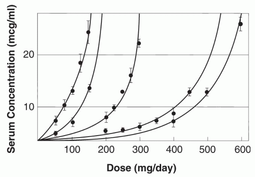
Enzyme saturation kinetics leads to phenytoin plasma concentrations increasing nonproportionally with changes in dose (Fig. 55.2) (75). The relationship between dose and concentration can be expressed by the Michaelis-Menten equation:

Dose (mg/day) = VmaxCss/Km + Css

where Vmax is the maximal rate of drug metabolism, Css the steady-state serum concentration, and Km the concentration
at which Vmax is half-maximal. The mean apparent phenytoin Km in adults 20 to 39 years old is 5.7 μg/mL (range, 1.5 to 20.7 μg/mL); the mean Vmax is 7.5 mg/kg/day (76). In most patients, phenytoin exhibits nonlinear pharmacokinetics because the usual therapeutic plasma concentrations exceed the usual Km. Concomitant illnesses (77) or medications, pregnancy (78), genetic makeup (79, 80, 81), and age can significantly affect Vmax or Km (or both). Children have higher Vmax values, but similar Km values, compared with adults (82, 83, 84); elderly individuals have lower Vmax values (mean, 6.0 mg/kg per day) (76).


Figure 55.2 Relationship between serum phenytoin concentration and daily dose in five patients. Each point represents the mean (± standard deviation [SD]) of three to eight measurements at steady state. The curves were fitted by computer through use of the Michaelis-Menten equation. (From Richens A, Dunlop A. Serum phenytoin levels in the management of epilepsy. Lancet 1975;2: 247-248, with permission.)


Excretion

Up to 95% of phenytoin is excreted in urine and feces as metabolites, with 5% or less of unchanged phenytoin excreted in urine. Phenytoin is also excreted in breast milk (85). Some investigators have suggested that phenytoin enhances its own elimination through enzyme induction (86).


Fosphenytoin


Absorption and Bioavailability

Fosphenytoin can be administered either intravenously or intramuscularly. The values for the area under the plasma total phenytoin and free phenytoin concentration versus time curves, after either intravenous or intramuscular administration of fosphenytoin, are almost identical to that for intravenous phenytoin sodium, indicating complete bioavailability by either route (11). These findings are based on studies involving single-dose intravenous and intramuscular administration to drug-free volunteers and single-dose intravenous administration to patients with therapeutic plasma phenytoin concentrations (11,87,88).

The total and complete conversion to phenytoin presents a potential clinical problem. A mg for mg PE conversion from oral phenytoin (Dilantin) capsules to parenteral fosphenytoin (Cerebyx) solution represents a 9% increase in total dosage, because 100-mg Dilantin capsules actually contain only 92 mg of phenytoin. Dosage adjustment is not usually necessary when Cerebyx is used for up to 1 week, although a phenytoin plasma concentration should be checked after longer periods of administration.


Distribution


Protein Binding

Like phenytoin, fosphenytoin is highly bound (95% to 99%) to serum albumin in a nonlinear fashion (11). This protein binding is not affected by prior diazepam administration (89). However, in the presence of fosphenytoin, phenytoin is displaced from binding sites, rapidly increasing unbound phenytoin concentrations as a function of plasma fosphenytoin concentration. This displacement is accentuated by fosphenytoin doses of at least 15 mg PE/kg delivered at rates of 50 to 150 mg PE per minute. As plasma fosphenytoin concentrations decline, phenytoin protein binding returns to normal. There is little displacement of phenytoin after intramuscular administration of fosphenytoin (11).


Volume of Distribution

Fosphenytoin’s volume of distribution is reported to be 0.13 L/kg in patients receiving 1200 mg PE fosphenytoin at 150 mg PE per minute. At lower doses and slower infusion rates, the volume of distribution is lower, 2.6 L, or approximately 0.04 L/kg for a 70-kg human (11,87,90). Fosphenytoin, a very polar molecule, achieves a rapid equilibrium between plasma and associated tissues (90).


Metabolism

After intravenous or intramuscular administration, the phosphate group of fosphenytoin is cleaved by ubiquitous nonspecific phosphatases to produce active phenytoin. The half-life of this conversion is approximately 8 to 18 minutes, is complete in a little more than an hour, and is independent of age, dose, or infusion rate (11,16,91, 92, 93). The tissue phosphatases responsible for this conversion are present at all ages; age, plasma phenytoin or fosphenytoin concentrations, and other medications do not alter their activity. The conversion of fosphenytoin to phenytoin is slightly faster in patients with hepatic or renal disease, consistent with decreased binding of fosphenytoin to plasma proteins and increased fraction of unbound fosphenytoin resulting from hypoproteinemia in these diseases (91). In addition, fosphenytoin’s phosphate load of 0.0037 mmol phosphate/mg PE fosphenytoin should be considered in patients with severe renal impairment (16).

A pharmacokinetic meta-analysis of plasma total and free phenytoin concentration from seven clinical trials involving neurosurgical patients, patients with status epilepticus,
patients with stroke, and healthy volunteers demonstrated that fosphenytoin loading doses of 15 to 20 mg PE/kg administered either intravenously or intramuscularly consistently resulted in total phenytoin plasma concentrations of 10 μg/mL or more and free phenytoin concentrations of 1 μg/mL or more. These therapeutic plasma phenytoin concentrations were reached in most subjects within 10 minutes, if rapid intravenous fosphenytoin dosing (≥100 mg PE per minute) was used, or within 30 minutes, if slower intravenous (<100 mg PE per minute) or intramuscular fosphenytoin dosing was used (94).


Figure 55.3 Free phenytoin concentration achieved in patients receiving an equivalent intravenous phenytoin loading dose (20 mg/kg) at 50 mg per minute and an equivalent intravenous fosphenytoin loading dose at 150 mg PE per minute. (From Eldon M, Loewen G, Voightman R, et al. Pharmacokinetics and tolerance of fosphenytoin and phenytoin administration intravenously to healthy subjects. Can J Neurol Sci 1993;20:5810, with permission.)

In one study, after administration of 1200 mg of phenytoin at 50 mg per minute, peak unbound phenytoin concentrations of approximately 3 μg/mL were achieved within 0.5 hour; administration of the equivalent fosphenytoin dose, infused at a rate of 150 mg PE per minute, produced similar peak unbound phenytoin concentrations (Fig. 55.3) (95). This rapid infusion rate was well tolerated. Therefore, when rapid achievement of therapeutic phenytoin concentrations is critical, as in the treatment of status epilepticus, fosphenytoin should be administered at a rate of 150 mg PE per minute. Slower infusion rates (50 to 100 mg PE per minute) may be acceptable in non-emergencies (95).


Excretion

A clinically insignificant amount of fosphenytoin (0 % to 4% of a dose) is excreted renally (93).


PLASMA DRUG CONCENTRATIONS


Phenytoin

Most laboratories and textbooks assume a therapeutic range for phenytoin of 10 to 20 μg/mL, which clinical experience and literature have called into question. Seizures have been controlled with concentrations lower than 10 μg/mL (96), although often, more than 20 μg/mL is needed. In one study (97), 51% of patients achieved complete control at concentrations either below or above that range. No significant association was evident between the serum phenytoin concentration and any measures of efficacy or toxicity.


Fosphenytoin

Measurement of fosphenytoin levels does not provide clinically useful information for patient care but rather has been utilized only in clinical research settings. Fosphenytoin may interfere with the ability of common laboratory immunoanalytic techniques, such as TDx/TDxFLx (fluorescence polarization) and Emit 2000 (enzyme multiplication), to measure phenytoin levels, because of cross-reactivity resulting in an artifactually elevated phenytoin concentration value. Waiting until all of the fosphenytoin to phenytoin conversion has occurred (approximately 2 hours after intravenous fosphenytoin administration, or 4 hours after intramuscular fosphenytoin administration) before attempting to measure a patient’s phenytoin concentrations is recommended (11).


DRUG INTERACTIONS


Phenytoin

Phenytoin can affect, and be affected by, a number of medications (Tables 55.2 and 55.3) (98). Although these drug interactions do not preclude concomitant administration, they signal the need for more frequent determination of serum concentrations, increased monitoring for the appearance of side effects, and, if appropriate, changes in dose. Patient-specific factors, such as genetic makeup, previous exposure to other compounds, and susceptibility to the clinical outcomes of the interaction, govern the extent and clinical significance of any drug interaction. In addition, a drug may act as an inhibitor in one patient and an inducer in another (e.g., phenobarbital’s effect on phenytoin).

Interactions can affect any of the four primary pharmacokinetic phases. A drug that affects absorption most likely will decrease phenytoin serum concentration. For example, administration of phenytoin with a continuous high-calorie, nitrogen liquid complete-nutrition formula through nasogastric tube feedings causes a decrease in phenytoin serum concentrations from a mean of 9.8 μg/mL to 2.72 μg/mL at the same dose (99).

Drugs that affect protein binding increase the percentage of unbound phenytoin, usually with no change in the unbound concentration and with a decrease in the total concentration. Valproic acid displaces phenytoin from protein-binding sites. When valproic acid is added to a phenytoin regimen, total phenytoin concentrations decrease, free
fraction increases, and free concentrations either stay the same or increase slightly. The following equation may be used to measure unbound phenytoin concentration in a patient receiving this combination (100,101):


TABLE 55.2 BIDIRECTIONAL INTERACTIONS BETWEEN PHENYTOIN AND OTHER ANTIEPILEPTIC DRUGS


Specific Drug


Effect of AED on Phenytoin Concentration


Mechanism of AED Effect


Effect of Phenytoin on AED Concentration


Mechanism of Phenytoin Effect


Carbamazepine


↑↓


CYP2C19 induction


↓↓


CYP3A4 induction


Ethosuximide


«


↓↓


CYP3A4 induction


Felbamate


↑↑


CYP2C19 inhibition


↓↓


CYP3A4 induction


Fosphenytoin


↑ Free phenytoin


Protein-binding displacement


«


Gabapentin


«


«


Lamotrigine


«


↓↓


UDPGT induction


Levetiracetam


«


«


Oxcarbazepine


CYP2C19 inhibition


↓ MHD


Unknown


CYP2C9 and


Phenobarbital


↑↓


CYP2C19 induction


Unclear


Topiramate


CYP2C19


↓↓


Unknown


Tiagabine


«


↓↓


CYP3A4 induction


Valproic acid


↓/ ? ↑ Free phenytoin


Protein-binding displacement and CYP2C9 inhibition


↓↓


CYP2C9 and CYP2C19 induction


Zonisamide


«


↓↓


CYP3A4 induction


↑↓, variable; ↑, minor increase; ↓, minor decrease; ↑↑, important increase; ↓↓, important decrease; «, no change; MHD, 10-monohydroxy derivative.


free PHT = [0.095 + 0.001(VPA)] PHT

where PHT = phenytoin and VPA = valproic acid.

Metabolic interactions usually cause either enzyme induction or inhibition. Addition of an inducer decreases phenytoin concentrations; addition of an inhibitor increases them. The order of addition or deletion is important. An inducer added to another compound may lead to decreases in the serum concentration of the preexisting drug; however, if that same drug is added to the inducer, the interaction would have a less noticeable clinical significance because nothing has changed—the added drug would simply require a higher dose. When an enzyme-inhibiting drug is removed from a regimen, the concentration of the remaining compound is likely to increase (102).

Only gold members can continue reading. Log In or Register to continue

Stay updated, free articles. Join our Telegram channel

Oct 17, 2016 | Posted by in NEUROLOGY | Comments Off on Phenytoin and Fosphenytoin

Full access? Get Clinical Tree

Get Clinical Tree app for offline access