2 Basic neuroanatomy and neurophysiology
At the end of this chapter you should be able to:
• identify the major subdivisions of the brain and the key brain areas contained within them
• identify the names and key functions of the 12 cranial nerves
• describe the structure of both the neurone and the synapse
• describe the events taking place during an action potential, with particular emphasis on the movement of sodium and potassium ions.
Introduction
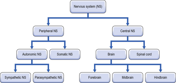
When trying to navigate around the complexities of the human nervous system, it is always a much less daunting prospect if you have a good map. Before we go on and look at the workings of the nervous system in the rest of the book, it is important that we find our bearings. The simplest way to subdivide the human nervous system is into central and peripheral sections. This, and subsequent divisions, can be seen in Figure 2.1.
As we advance through this book we shall deal with various areas of the brain and it is important that you have a clear understanding of at least the simplest level of brain anatomy. The three primary brain regions (forebrain, midbrain and hindbrain) can be further sub-divided as shown in Table 2.1.
Table 2.1 Anatomical subdivisions of the brain
| Forebrain | Telencephalon | Cerebral cortex |
| Basal ganglia | ||
| Limbic system | ||
| Diencephalon | Thalamus | |
| Hypothalamus | ||
| Midbrain | Mesencephalon | Tectum |
| Tegmentum | ||
| Hindbrain | Metencephalon | Cerebellum |
| Pons | ||
| Myelencephalon | Medulla |
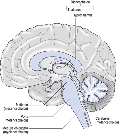
In order to fully appreciate the orientation of the brain it is often useful to imagine a sagittal section through the brain from front to back. This results in a view of the inner and cortical surfaces, as seen in Figure 2.2.
The forebrain
The telencephalon comprises the majority of the two cerebral hemispheres that make up the cerebrum. The top, visible surface of the cerebral hemispheres is the cerebral cortex which sits on top of the sub-cortical structures of the limbic system and the basal ganglia. The name cortex derives from the Latin word for the bark of a tree. The cerebral cortex is about 3 mm thick, covers the two hemispheres of the brain and has a ridged surface comprised of grooves (sulci) and peaks (gyri). The larger grooves that can be seen on the cortical surface are the fissures. This groove/peak structure significantly increases the surface area of the cortex, taking it up to approximately 0.25 m2 (2500 cm2). The cortex can be further subdivided into four lobes: frontal, occipital, parietal and temporal (Fig. 2.3) and the location of cortical structures are often identified in relation to the various lobes.
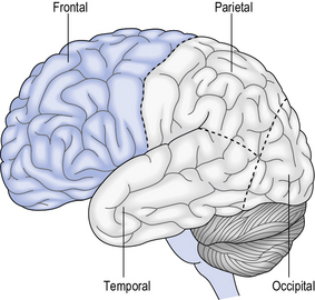
Fig. 2.3 • External appearance of the brain, showing the lobes.
From Kindlen (2003) with permission from Elsevier
Basal ganglia
The basal ganglia are a collection of subcortical nuclei consisting of the caudate nucleus, the putamen, the nucleus accumbens, the substantia nigra and the globus pallidus. The primary function of these interconnected nuclei is an involvement in the control of movement. A better understanding of the role played by the basal ganglia can be achieved when there is a degeneration of specific neurones in the midbrain that project to the caudate and the putamen (i.e. the nigrostriatal dopaminergic pathway). This form of degeneration results in Parkinson’s disease (PD), the symptoms of which include limb rigidity, poor balance, muscle weakness, tremor and a difficulty in initiating movements. Chapter 6 will explain the function of the basal ganglia in more depth.


