Basic Overview of Electromyography

12 Basic Overview of Electromyography


After the nerve conduction studies are completed, the electrophysiologic evaluation moves on to the needle electromyography (EMG) examination. Like the nerve conduction studies, each needle EMG study must be individualized based on the clinical findings and differential diagnosis and modified as the test proceeds and more data are obtained. Almost every muscle in the body can be studied with EMG. However, to do so is neither practical for the electromyographer nor desirable for the patient. For each study, a balance must be reached between the need to study a sufficient number of muscles to reach or exclude a diagnosis and the limits of the patient’s ability to tolerate the examination. Patients’ reactions to the needle EMG vary greatly. When the examination is performed skillfully, most patients tolerate it well, with only minor discomfort. Some patients, however, are extremely apprehensive and may have difficulty completing the examination. Young children, who may tolerate the nerve conductions well, frequently have difficulty with the needle examination. It is with these latter groups that the electromyographer must be especially skillful. Before proceeding with the needle study, it often is useful to consider the possibility that the patient may tolerate EMG of only one or two muscles. If this occurs, which muscles will one choose? The choice must be based on the following factors:


If there is any indication that the patient might not tolerate or complete the entire examination, the most important muscles should be sampled first. For instance, if a patient has proximal muscle weakness and the differential diagnosis rests primarily between a myopathy and a proximal neuropathic process (e.g., plexopathy, radiculopathy, motor neuron disease), it makes sense to sample a weak proximal muscle first. If one begins the examination by sampling distal muscles that are clinically normal and the patient asks to stop the examination after the distal muscles are sampled, the chance to reach a diagnosis may have been lost.


There is no doubt that the needle EMG is the more challenging part of the electrophysiologic examination. A successful study requires not only knowledge of anatomy and physiology but also sound EMG technique and good patient rapport. Two competing influences make the needle EMG study especially demanding. First, many of the abnormalities found on the needle study are subtle. At the same time, however, the range of normal findings is quite large and varies with age and with the muscle being studied. Although the basics of the needle EMG study, such as needle placement and recognition of certain types of abnormal spontaneous activity, usually can be learned in a short time, it is not unusual for it to take years to master recognition of many of the uncommon and subtle needle EMG findings.


Equipment


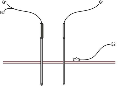
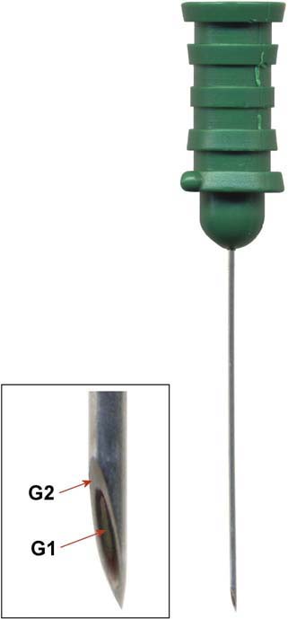
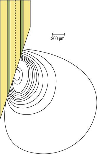
In addition to the EMG machine, an EMG needle, needle cable, ground electrode, and gloves are necessary to perform the needle EMG study. The ground electrode is applied to the limb being studied in order to suppress noise and for electrical safety. Disposable gloves must always be worn to prevent the transmission of bloodborne infections between the patient and the electromyographer. The EMG needle is connected to a cable and then plugged into the EMG machine. Either a concentric or monopolar EMG needle can be used (Figure 12–1). When an electrical potential is measured, including the potentials measured during the needle EMG study, voltage is measured as the difference between the active and reference recording electrodes. The concentric needle contains both the active and reference electrodes in the needle itself (Figure 12–2). The shaft of the needle serves as the reference electrode, whereas the active electrode runs as a very small wire through the center of the needle and is exposed at the needle tip. The end of the concentric needle is beveled, resulting in a recording area that has a “teardrop” configuration (Figure 12–3). In contrast, the monopolar needle is Teflon coated, and its exposed end serves as the active recording electrode. Its recording area is that of a sphere around the tip of the needle. For the monopolar needle montage, an additional surface disc electrode is required as the reference electrode.


Stay updated, free articles. Join our Telegram channel

Aug 31, 2016 | Posted by in NEUROLOGY | Comments Off on Basic Overview of Electromyography

Full access? Get Clinical Tree

Get Clinical Tree app for offline access