Biologic Therapy for Disk Degeneration


Biologic Therapy for Disk Degeneration


Daniel K. Park

Michael B. Ellman

Howard S. An


Low back pain (LBP) associated with degenerative disk disease is a common clinical problem that has a tremendous socioeconomic impact in today’s aging population. Economic losses have been estimated to exceed 90 billion dollars per year (1) as eight of every ten adults will, at some point, suffer from LBP that impairs activities of daily living (2). Furthermore, about two-thirds of people will experience neck pain during their life, up to 45% each year (3,4). The prevalence is highest in middleaged patients, women more common than men (5). In some industrial countries, neck-related pain account for as much time off work as LBP (6). Despite the significant impact, the exact pathophysiology of LBP and neck pain is not yet understood. Intervertebral disk (IVD) degeneration has been associated with spinal pain, but a direct correlation has not been confirmed. Scientific research has demonstrated that progressive degeneration of the spinal extracellular matrix (ECM) is closely associated with symptomatology. Therefore, ECM degeneration may play a pivotal role in the pathophysiology of this clinical entity.


INTERVERTEBRAL DISK STRUCTURE

The anatomy of the IVD must be understood prior to exploring various biologic therapies aimed at reversing disk degeneration and providing symptomatic relief of LBP. The IVD is composed of two layers. An outer fibrous layer, the annulus fibrosis (AF), provides tensile strength, while the hydrostatic properties of the inner nucleus pulposus (NP) provide compressive strength (7, 8 and 9).

Within these two substructures, the interactions of two proteins, proteoglycans (PG) and collagen, allow for proper biomechanical function (10). In the inner NP, notochordal cells and disk cells synthesize PGs. The hydrophilic nature of the PG attracts and holds water, providing the compressive cushion of the disk. In contrast to PGs, collagens provide tensile strength to the IVD (11,12). Collagen types I and II make up 80% of the I VD. Type I is mainly located in the AF while type II is predominantly found in the NP. Further, the collagen fibers of the outer AF blend with the posterior longitudinal ligament inserting into the vertebral bodies, allowing tensile hoop stresses to be transferred to the outer AF and permitting IVD motion (13,14).


DISK DEGENERATION

Degeneration of the spine is an inevitable consequence of aging. Miller and colleagues found that disk degeneration occurs in roughly 16% of the population early in life and approximately 98% later in life, based on macroscopic disk degeneration grades (15). Although spinal degeneration is inevitable with aging, it is typically asymptomatic (16,17). The diagnostic dilemma, therefore, is discerning whether degenerative disks are a mere consequence of aging or are pathologic and symptomatic in nature.

Various factors have been implicated in the etiology of disk degeneration (18,19). One factor is decreased IVD nutrition (20,21). The blood vessels that supply the center of the disk disappear by the third decade of life, resulting in the disk relying only on diffusion through the end plate for survival (22). In adults, cells in the center of the NP may be 7 to 8 mm from the nearest blood supply and receive little, if any, diffusion of nutrients from end plate vasculature (22). The resultant decrease in oxygen tension ultimately results in anaerobic metabolism and the production of lactic acid. Lactic acid production subsequently creates a more acidic-surrounding environment, which negatively affects cell viability (20) and hinders ECM production in the NP (23).

In addition to a decrease in nutritional support, cell viability (20,24,25) and synthetic capacity of surviving cells (26) are suppressed with IVD aging and degeneration. Histochemical studies have found that in human surgical disks with degeneration, a high incidence of cellular apoptosis is present and the surviving cells are not synthetically inactive but rather produce inappropriate matrix products (27). Furthermore, while collagen content remains relatively stable over time, collagen
properties such as solubility and mechanical strength are altered because cross-links are formed nonenzymatically, as compared to enzymatically mediated pyridinoline cross-linking found in young, healthy disks (28,29). Compared to the relatively constant collagen content, PG content in the NP decreases noticeably with age and degeneration (30, 31, 32 and 33), ultimately diminishing the waterholding capacity of the IVD and reducing its ability to distribute loads effectively. In the AF, however, the water content remains relatively constant, and chemical mediators may play a more significant role in annulus degeneration (34). Importantly, enzymatic by-products of degeneration, such as fibronectin and aggrecanase fragments, may further promote the degeneration process in both NP and AF tissues(35,36).

Another factor implicated in disk degeneration involves biomechanical processes secondary to alterations in mechanical loading (12,37). One hypothesis is that the mechanical environment in aging and/or degeneration produces localized trauma to the disk. Because of the slow turnover of the disk cells, repair will be minimal (38). Accumulation of these “microtraumas” over time, accompanied by the decreased turnover and synthetic rate of disk cells and matrix, ultimately leads to progressive weakening of the disk and degeneration. In addition, prolonged mechanical loading causes a higher rate of disk cell apoptosis (39). Thus, as subtle instability occurs within the vertebral spinal units, changes in biomechanical loading can ultimately result in biochemical changes leading to cell death and IVD degeneration.

Lastly, genetic factors have been proposed as a cause of disk degeneration. Battie et al. (40) reported that genetic factors may play a primary role in disk degeneration in identical twins. Videman et al. (41) were the first to describe that specific genes, specifically the vitamin D receptor gene, were associated with disk degeneration. Other possible genetic associations include aggrecan gene polymorphism (42), interleukin-1 (IL-1) and COL9A3 (43,44), COL9A2 (45), collagen IX and XI (46), and matrix metalloproteinase (MMP)-3 (47).

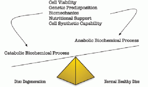
Given the multiple and complex etiologies leading to disk degeneration (Fig. 99.1), it may be easier to focus therapeutic strategies on preventing the ultimate outcome: biologic NP and AF degradation. On a biologic level, the etiologies outlined above result in degeneration of the IVD, which stems from an imbalance between anabolic and catabolic biochemical processes (48). This delicate balance is normally maintained by disk cells residing in both the AF and NP, actively regulating IVD homeostasis through a variety of stimuli, including cytokines, enzymes, enzyme inhibitors, and growth factors (49,50). In degenerative disks, at least in vitro, an increase in the expression of proinflammatory cytokines and growth factors, coupled with the gradual loss of large PGs such as aggrecan from the NP, have been reported (51). Linked to these changes is an increased expression of matrixdegrading enzymes such as MMPs and aggrecanases (a disintegrin and metalloprotease with thrombospondin motifs), both of which are produced by native cells (51). Consequently, matrix homeostasis favors a procatabolic state, leading to ECM degradation and further disk degeneration (Fig. 99.1).


Figure 99.1. Factors that influence disk degeneration. Multiple factors influence the delicate balance of disk degeneration and the maintenance of healthy disks.


PREVENTION OF DISK DEGENERATION USING BIOLOGIC FACTORS

While alterations in both anabolic and catabolic processes are thought to play key roles in the onset and progression of IVD degeneration, the biochemical processes that regulate these changes are still poorly understood. One strategy to retard the progression of disk degeneration and to ameliorate the loss of structural integrity is to shift the metabolic status from catabolic to actively anabolic by stimulating disk cells with biologic factors. For example, the upregulation of important matrix proteins such as aggrecan, along with a downregulation of proinflammatory cytokines and matrix-degrading enzymes, may serve to inhibit and/or reverse disk degeneration, and biologic factors mediating these activities serve as potential therapeutic strategies to treat and/or prevent disk degeneration (52).

Clinically, the application of biologic factors to treat degenerative disk disease has significant potential, yet a detailed analysis of their mode of action is necessary for several reasons. Both in vitro and in vivo studies are critical to understand indication, optimal dose, mode of delivery, duration of action, and side effects of each potential therapy. Biologic factors may have a different effect on the NP compared to the AF or exert different actions at different stages of degeneration (53,54). For example, cells found in a disk with an advanced stage of degeneration may not respond to factor-induced stimulation, while those cells at an earlier stage may respond well. Thus, in vitro and in vivo studies are essential to explore the therapeutic use of biologic factors on IVD degeneration.

Several authors have identified biologic factors involved in the stimulation of PG synthesis, inhibition of catabolic enzymes, and restoration of disk height in animal models of the IVD (49,50,55, 56, 57, 58, 59, 60 and 61). Thompson et al. (55) were the first to elucidate the effects of anabolic growth factors on the IVD. The authors used a canine IVD organ culture system to investigate the effects of insulin-like growth factor (IGF-1), epidermal growth factor (EGF), fibroblast growth factor (FGF), and transforming growth factor-β (TGF-β) on IVD PG synthesis and cell proliferation. The authors reported anabolic effects of TGF-β, EGF, and basic FGF (bFGF) on
PG synthesis, with more pronounced effects in the NP compared to AF. Since the Thompson et al. (55) landmark report, several studies have demonstrated the importance of biologic factors in IVD homeostasis (49,50) (Table 99.1). Below, we review several pertinent biologic factors that have been shown to slow or reverse disk degeneration in vitro and/or in vivo, elucidating the significant potential for clinical application of biologic factors in the future.


OSTEOGENIC PROTEIN-1

Osteogenic protein-1 (OP-1, otherwise known as bone morphogenetic protein-7) is expressed in cartilage and was initially found to exert potent anabolic effects on osteocyte and chondrocyte differentiation and metabolism (57,62). Recently, the anabolic effect of OP-1 on cartilage regeneration in both articular cartilage and spine disks has been elucidated. OP-1 has been shown to stimulate PG and collagen synthesis in human adult articular chondrocytes (63). In the IVD, Takegami et al.(64) and An et al. (58) described the anabolic effects of OP-1 via increased PG synthesis of rabbit disk cells from the NP and AF when treated with OP-1 in vitro. Masuda et al. (59) first reported the stimulatory effect of OP-1 on PG and collagen metabolism in rabbit NP and AF cells cultured in alginate beads. In both cell types, recombinant human OP-1 (rhOP-1) stimulated the
synthesis of PGs and collagens in a dose-dependent manner with an associated increase in the expression of mRNA for aggrecan and collagen type II. In addition, continuous treatment with rhOP-1 stimulated the accumulation of ECM as well as an increase in cell number, suggesting that the stimulation of matrix synthesis is associated with the retention of newly synthesized matrix molecules. Although data suggest that the capacity of NP and AF cells to respond to growth factors diminishes with age (61), it has been found that the responsiveness of IVD cells to OP-1 is independent of age (60). Further, Imai et al. (65,66) reported that OP-1 enhanced the in vitro production of PG by human NP and AF cells cultured in alginate beads. Similar to rabbit cells, OP-1 also enhanced the accumulation of PG in the matrix. Interestingly, AF cells, which are more fibrochondrocytic than NP cells, strongly responded to OP-1, suggesting that OP-1 might be beneficial not only for nucleus repair but for annulus repair as well (65).


TABLE 99.1 In Vitro and In Vivo Studies of the Effects of Biologic Factors on IVD Homeostasis


Factor


Cell Type


Effect


Culture Type


References


OP-1


Human NP, AF


PG accumulation ↑, ECM production


Alginate bead


(61)


OP-1


Rabbit NP, AF


PG synthesis ↑, cell proliferation ↑, aggrecan, collagen II mRNA ↑


Alginate bead


(55,60)


OP-1


Rabbit NP, AF


After IL-1 tx, PG accumulation ↑, ECM production ↑


Alginate bead


(63)


OP-1


Rabbit NP, AF


After C-ABC tx, PG accumulation ↑, ECM production ↑


Alginate bead


(60)


OP-1


Rabbit IVD


OP-1 injections ↑ disk height, initial PG content in NP↑


In vivo compression model


(54)


OP-1


Rabbit IVD


After needle puncture-induced disk degeneration, OP-1 ↑ disk height, ↑ signal intensity (T2-MRI), ↑ PG content


In vivo


(53,54)


OP-1


Rabbit IVD


After chemonucleolysis degeneration by C-ABC injection, OP-1 ↑ disk height


In vivo


(62,65)


OP-1


Rabbit IVD


After coinjection with C-ABC, OP-1 ↑ disk height, initial PG content in NP ↑


In vivo


(66)


BMP-2


Human NP, TZ, degen disk


PG synthesis ↑, cell proliferation ↑, aggrecan, collagen I & II mRNA ↑


Monolayer


(68)


BMP-2


Rat AF&TZ


ECM ↑, PG synthesis ↑, cell proliferation ↑, type II collagen, aggrecan and Sox9 mRNA→


Monolayer


(69)


BMP-2


Rabbit IVD with annular tears


BMP-2 injection ↑ fibroblast proliferation, ↑ vascularity after injury, role in healing


In vivo


(52)


TGF-β


Canine NP, AF, TZ


PG synthesis ↑, cell proliferation ↑ (NP, TZ)


Organ culture


(51)


TGF-β


Human AF


↑ cell proliferation, ↑ PG synthesis


Monolayer, alginate, agarose


(74)


GDF-5


Rat IVD


Injection of GDF-5 ↑ disk height, clustering of cells


In vivo compression model


(80)


RSV


Bovine NP, AF


PG synthesis?, PG accumulation ↑, suppress catabolic effects of bFGF and IL-1


Alginate, monolayer


(88)


IL-1ra


Human NP, AF


Gene transfer of IL-1ra ↑ levels of IL-1ra and suppresses catabolic effects of IL-1


Gene transfer


(92)


PDGF


Canine NP, AF, TZ


No effect


Organ culture


(51)


PDGF


Human AF


Apoptosis ↓ in apoptotic-induced environment


Monolayer


(23)


IGF-1


Bovine NP, AF


PG synthesis ↑, cell proliferation ↑


Monolayer


(96)


IGF-1


Human AF


Apoptosis ↓ in apoptotic-induced environment


Monolayer


(23)


IGF-1


Canine NP, AF, TZ


PG synthesis ↑, cell proliferation ↑


Organ culture


(51)


EGF


Canine NP, AF, TZ


ECM ↑, PG synthesis ↑, cell proliferation ↑


Organ culture


(51)


bFGF


Canine NP, AF, TZ


PG synthesis ↑


Organ culture


(51)


bFGF


Rat and human NP, AF


Cell proliferation ↑


Organ culture


(97,98)


bFGF


Bovine NP, AF


Matrix-degrading enzyme expression ↑, PG synthesis ↓, suppresses action of OP-1


Monolayer, alginate


(99)


bFGF


Ovine AF


bFGF ↑ over time after disk injury, cell proliferation ↑


Immunohistochemistry


(105)


OP-1, osteogenic protein-1; BMP-2, bone morphogenetic protein-2; TGF- β, transforming growth factor- β; GDF-5, growth differentiation factor-5; RSV, resveratrol; IL-1ra, interleukin-1 receptor antagonist; PDGF, platelet-derived growth factor; IGF-1, insulin-like growth factor; EGF, epidermal growth factor; bFGF, basic fibroblast growth factor; AF, annulus fibrosus; NP, nucleus pulposus; TZ, transitional zone; PG, proteoglycan; ECM, extracellular matrix; Tx, treatment; C-ABC, chondroitinase-ABC; IVD, intervertebral disk.

Only gold members can continue reading. Log In or Register to continue

Stay updated, free articles. Join our Telegram channel

Jul 5, 2016 | Posted by in NEUROSURGERY | Comments Off on Biologic Therapy for Disk Degeneration

Full access? Get Clinical Tree

Get Clinical Tree app for offline access