CHAPTER 98 Brain Tumor Stem Cells
Brain tumor stem cells (BTSCs), a small subset of cells found within multiple types of central nervous system (CNS) malignancies, possess two unique qualities that distinguish them from other cells in the tumor stroma: (1) they have all the properties needed to fulfill the criteria for defining stem cells, namely, the ability to self-renew and to differentiate into the three lineages of nervous tissue (astrocytes, oligodendrocytes, and neurons) under appropriate conditions and (2) they have the ability, on injection into the brain of non–immune-competent animals, to initiate brain tumors that recapitulate the growth and infiltrative patterns of the tumor of origin.1,2 The BTSC hypothesis is dependent on the idea that there exists a pool of cells in all brain tumors that contain the necessary genetic programming for tumorigenesis, whereas the remaining cells serve as non–tumor-initiating constituents of the malignant tissue.3 The origin of these BTSCs has yet to be clearly identified, with some implicating abnormal transformation of naturally occurring neural stem cells (NSCs) as the primary source.2 Others speculate that more dedicated cells along the path of differentiation can “revert” and initiate the critical functions of self-regeneration and aberrant proliferation.4 The BTSC hypothesis has some important clinical implications: cancer-initiating stem cells exhibit strong migratory behavior5 and are exquisitely radioresistant and chemoresistant.6 This may serve to explain the local metastatic potential and resistance to treatment of the majority of malignant brain tumors. Rather than attempting to destroy the bulk of the tumor with the antineoplastic agents available today, some research groups have begun to focus on finding a method of selectively targeting this small, yet deadly, pool of tumorigenic BTSCs. Given the numerous similarities existing between normal stem cells and BTSCs, we can potentially exploit the bulk of knowledge about normal stem cell biology to identify possible novel molecular targets that could arrest BTSC proliferation.2 Some examples of therapeutic modalities include inhibitors of the sonic hedgehog (SHH) pathway (required for the proliferation of stem cells),7 induction of terminal differentiation of BTSCs with bone morphogenetic protein [BMP]),8 and disruption of the critical angiogenic niche of BTSCs.9 This chapter primarily reviews the concept of the neurosphere assay, evolution and development of the BTSC hypothesis, basic phenotypic properties of BTSCs, the role of molecular markers implicated in isolating these cells, the intracellular pathways used to maintain “stemness,” their relationship to NSCs, and finally, the clinical application of this hypothesis.
The Neurosphere Assay and Discovery of Adult Neurogenesis
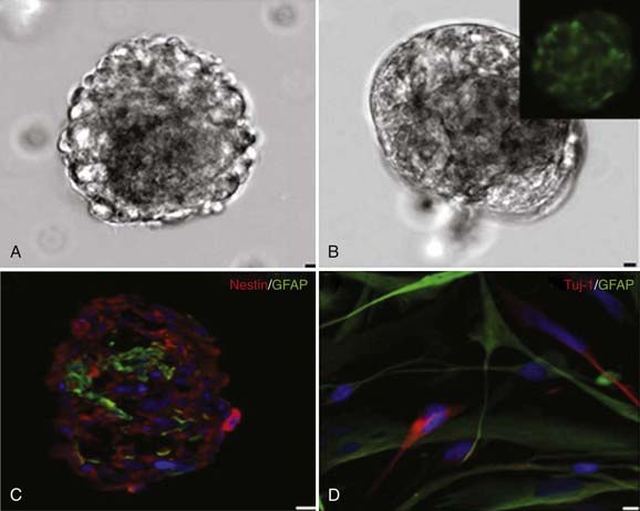
Advent of the neurosphere assay, originally developed for the isolation of adult NSCs, was critical to the identification of cancer-initiating stem cells from human brain tumors. It was originally believed that the adult brain did not retain the ability to produce new neurons in the postnatal period but was rather a static organ.2,10 Altman was the first to challenge this view when he used tritiated thymidine to monitor dividing cells in restricted areas of the neonatal rat brain in 1962.11 Altman was able to show that these dividing cells eventually differentiated into cells that morphologically resembled neurons.11 Kaplan and Hinds confirmed these observations in the 1970s by using electron microscopy to show that the postnatally dividing cells in the brain contained dendrites and axons.12 Introduction of the neurosphere assay, developed by Reynolds and Weiss, allowed in vitro isolation of normal stem cells from adult and fetal brain tissue (Fig. 98-1).13 In this assay, brain tissue is dissociated into single cells and cultured in special media containing various mitogens (i.e., basic fibroblast growth factor, epidermal growth factor).14 Under these conditions, NSCs form a ball of cells called “neurospheres” that contain the parent NSC and its differentiated progeny: neurons, oligodendrocytes, and astrocytes. The self-renewal capacity of these NSCs is confirmed by dissociating the neurospheres and plating individual cells per well with neurosphere media. The neurosphere-forming ability of each cell from the original neurosphere is then assessed to determine “stemness.” This assay allowed identification and isolation of adult NSCs in the subventricular zone (SVZ) of the lateral ventricles and the subgranular layer of the hippocampal dentate gyrus.13,15
It is now recognized that there is a strict hierarchy in the largest of these germinal centers, the SVZ, where adult stem cells systematically differentiate into more dedicated progenitor cells.16 NSCs are found as quiescent type B cells that give rise to the surrounding and more rapidly dividing type C cells. These cells subsequently differentiate into type A cells (neuroblasts), which surround the ventricles, separated only by a single layer of ependymal cells, and directly divide to form new neurons in the adult brain.17 Adult neurogenesis has been implicated in the formation of new neuronal connections and contributes to memory formation in the normal adult brain. More essential for our discussion, adult NSCs are also important because they are believed to undergo malignant transformation into BTSCs.18
Development of the Brain Tumor Stem Cell Hypothesis
For decades, the traditional belief in oncology maintained that human tumors represented a collection of cells in which a majority (or all) had the intrinsic ability to initiate tumorigenesis. The concept that only a small fraction of cells was ultimately responsible for tumorigenesis was originally proposed in 1963, when Bruce and Van Der Gaag recognized the ability of a small number of lymphoma cells to rapidly proliferate and differentiate in vivo.19 The theory was not supported until the advent of more sophisticated research tools that allowed scientists to recognize that not only were leukemia cells clonal in origin20 but also that cancer cells maintained a hierarchical organization similar to embryonic tissues, with only a small subset of proliferating progenitors replenishing the nonregenerating leukemia cells.21,22 This concept quickly spread to other types of malignancies, where it was noted that only a subset of cells from human breast,23 pancreas,24 prostate,25 head and neck,26 and colon27 cancer tissue had the ability to initiate tumorigenesis on transplantation.
Ignatova and colleagues described the isolation of neurosphere-forming, bipotent (neuronal/astroglial) precursors from glioblastoma multiforme (GBM).28 This was subsequently confirmed and extended by Singh and coworkers, who also demonstrated that bipotent lineage-restricted progenitors from CNS tumors display short-term self-renewal (three passages in culture).18 Similar findings were reported by Hemmati and coauthors, who described the absence of oligodendrocytes in these cells and also addressed the issue of tumorigenicity by demonstrating tumor formation after intracerebral transplantation of tumor-derived cells.29
Galli and colleagues took these simple observations further by showing that neurosphere-forming cells could also act as cancer-initiating cells and can establish tumors that closely resemble the main histologic, cytologic, and architectural features of the human disease even after multiple serial transplantations.30 This was later confirmed by Singh and associates with a different method of isolating BTSCs (using the NSC marker CD133, to be discussed later).31 Unlike NSC neurospheres, however, BTSC neurospheres consisted of abnormally differentiated cells with a genetic profile similar to that of the original tumor and a significantly increased proliferation rate.18,28–30,32,33 The number of in vitro neurospheres formed in a particular sample of human brain cancer was shown to be correlated with the more rapidly growing, aggressive tumor in vivo, thus suggesting that a poorer clinical outcome was associated with a greater number of brain tumor–perpetuating stem cells.34 These experiments suggested that not all cells within brain tumors had the ability to initiate tumorigenesis.18,29–31,35 Rather, brain tumors represented a hierarchy of malignant cells, with stem cells at the top of the hierarchy “differentiating” into the aberrant tumor stromal cells at the bottom. This basic organization was similar to normal adult neurogenesis, with a small colony of NSCs replenishing the cells of the SVZ11,12 and hippocampus.36 If these BTSCs were “differentiating” into malignant tumor stromal cells, it could be speculated that these less dedicated cells along the path of differentiation must express cell surface markers that vary from their progeny.32 This is supported by recent evidence suggesting that after multiple in vitro passages of primary GBM, the resulting cells are the product of an outgrowth of cell clones that have accumulated profound “de novo” genetic or epigenetic changes, or both. These clones do not share the same genetic profile or growth characteristics of the parent tumor.32
Markers for Neural Stem Cells and Brain Tumor Stem Cells
Nestin is an intermediate filament protein that is produced in both NSCs and progenitor cells during development.37 This molecule has been implicated as important in the morphology and adhesive properties of NSCs and is dramatically downregulated when NSCs differentiate into more committed lines. Interestingly, this marker is upregulated in brain tissue during times of injury, ischemia, and inflammation, which suggests that nestin-labeled cells may be responsible for remodeling of adult brain tissue after damage.38–40 Nestin has also been identified in a variety of human brain tumors, including ependymomas, malignant astrocytomas, oligodendrogliomas,41 and malignant gliomas.42 Nestin-positive cells from multiple brain cancer types have been shown to have significantly enhanced migration and invasive properties43 and readily form neurospheres. More importantly, this marker is a potential candidate for assessing malignant potential, with clinically malignant tissues exhibiting enhanced nestin expression.42 These results suggest that the number of proliferating nestin-labeled cells, or BTSCs, may predict a patient’s clinical outcome.
CD133 (prominin-1) is a cell membrane glycoprotein originally identified on primitive hematopoietic stem cells. It is a five-transmembrane protein that contains two large extracellular loops; it is found on various types of stem cells and known to be downregulated on differentiated cells.44,45 This marker was later identified in murine postnatal brain tissue,45 where cells sorted selectively for CD133 were able to both form neurospheres in vitro and differentiate into both neuronal and glial cell types, thereby fulfilling the definition of an NSC. Although its natural function is unknown, some speculate that CD133 has a role in the dynamic organization of cell surface protrusions. There is mounting evidence suggesting that adult NSCs and BTSCs share this cell surface marker. Singh and coworkers were the first to show that like adult NSCs, CD133-positive cells isolated from human glioblastomas and human medulloblastomas were able to form neurospheres in vitro.31 As few as 100 CD133-positive brain tumor–derived cells were necessary to successfully form tumors in immunodeficient mice, whereas more than 105 CD133-negative cells failed to drive tumor formation.31 Although this evidence points toward CD133 as a putative marker for BTSCs, more recent evidence has complicated the issue. Wang and coauthors reported that CD133-negative cells are also able to form tumors and give rise to CD133-positive cells, which correlated with poor survival in rat models.46 There exists a considerable amount of genetic heterogeneity in brain tumors, and it is likely that there may be multiple convergent pathways leading to the formation of brain tumors. As observed in leukemias and bone marrow stem cells, multiple markers are probably necessary to adequately characterize BTSCs.46,47
Cell of Origin of Brain Tumor Stem Cells
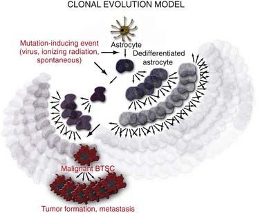
There are three possible explanations for the origins of BTSCs. The traditional clonal evolution hypothesis (Fig. 98-2) argues that brain tumors arise from dedifferentiation of dedicated adult brain cells.48 The subsequent clonal expansion of this single dedifferentiated cell and the accumulation of mutations over time confer the progeny with the ability to proliferate and regenerate in an unregulated manner. This process results in eventual formation of the BTSC, which produces the primary tumor focus and migrates locally to cause metastatic disease. With the discovery of adult NSC populations and the observation that BTSCs behave much like adult NSCs in terms of organization, proliferation, and regeneration, it was hypothesized that abnormal differentiation of adult NSCs may be the direct source of human brain tumors (Fig. 98-3).49 Finally, the progenitor recruitment hypothesis argues that a single tumor progenitor cell of undefined origin has the ability to recruit normally quiescent neural progenitor cells and induce them to proliferate in an unregulated manner via the release of local growth factors (Fig. 98-4). Definitive proof has yet to be found regarding which hypothesis is dominant in the natural pathogenesis of human brain tumors, and there is significant experimental evidence to support all three alternatives.
Stay updated, free articles. Join our Telegram channel

Full access? Get Clinical Tree


