Fig. 59.1
The progressive growth pattern of cavernous and clinoid segment internal carotid artery (ICA) (infradiaphragmatic) aneurysms (a–c)medially into the sella, causing compression of the pituitary gland (Courtesy of Dr. Arthur L. Day)
Supradiaphragmatic aneurysms, which arise from the ophthalmic segment of the ICA at the origin of the superior hypophysial artery, or anterior communicating artery aneurysms that point inferiorly into the sella (Fig. 59.2). These typically tend to be larger aneurysms (mean diameter 21 mm), and they are more likely to present with visual loss.
Fig. 59.2
The progressive growth pattern of superior hypophysial/ophthalmic ICA aneurysms (a–c)medially and intradurally above the diaphragma sellae, from where they may extend down into the sella turcica (Courtesy of Dr. Arthur L. Day)
Cranial nerve paresis is noted in 18 % of patients with intrasellar aneurysms [4].
Rarely, bilateral ICA aneurysms may impinge on the pituitary gland and cause various endocrinopathies [7, 8].
59.2 Imaging Features
Diagnosis of a sellar region aneurysm can be made with CT angiography, MR angiography, or conventional digital subtraction angiography (Figs. 59.3, 59.4, 59.5, 59.6, and 59.7).
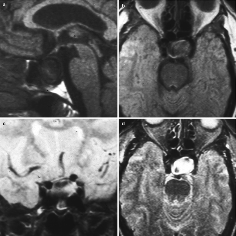
Fig. 59.3
Infradiaphragmatic sellar aneurysm. Sagittal T1-weighted (a), axial T1-weighted (b), coronal T2-weighted (c), and axial T2-weighted (d) MR images obtained in a patient with a left cavernous segment infradiaphragmatic aneurysm extending into the sella turcica (Reproduced with permission from Hanak et al. [4])
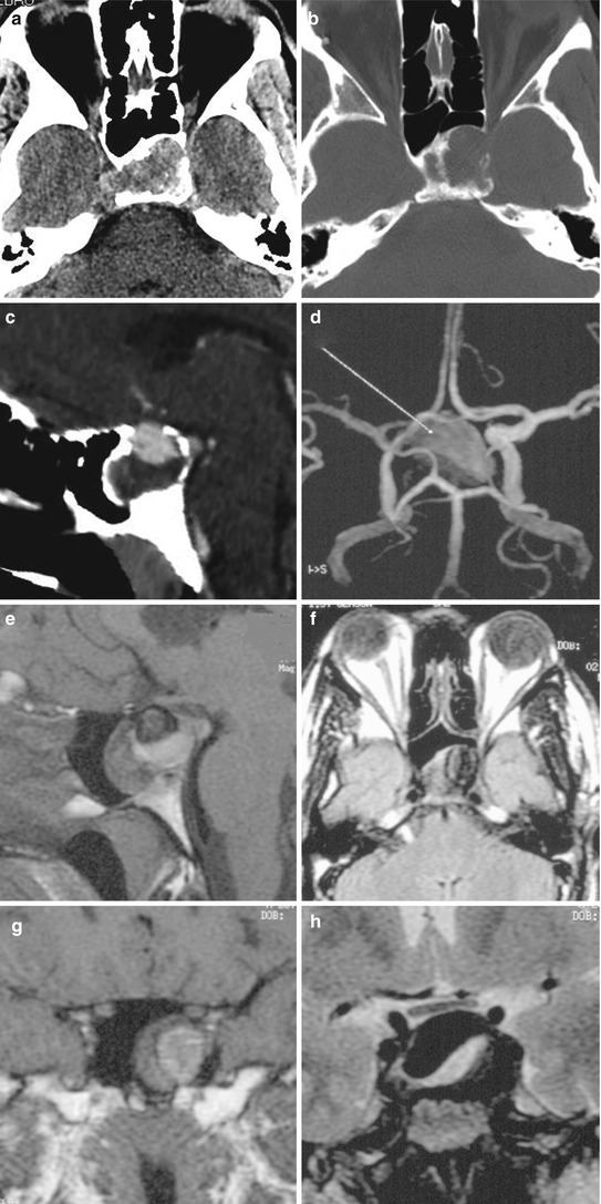
Fig. 59.4
Infradiaphragmatic sellar aneurysm. (a, b) Axial CT brain and bone windows obtained in a patient with a large, partially calcified cavernous ICA infradiaphragmatic aneurysm extending into the sella and sphenoid sinus. (c, d) Sagittal and 3D reconstructed CT angiograms demonstrating that the aneurysm is partially thrombosed. (e, f) Sagittal and axial T1-weighted postcontrast MR images also demonstrating partial thrombosis of this aneurysm. (g, h) Coronal T1-weighted postcontrast and T2-weighted MR images demonstrating extension into the sphenoid sinus and a marble-like appearance caused by the partial thrombosis (Reproduced with permission from Hanak et al. [4])
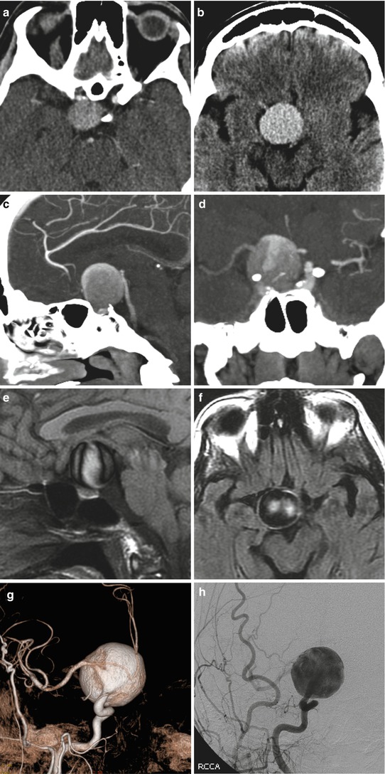
Fig. 59.5
Sellar aneurysm. (a, b) Axial CT scans obtained in a patient with a large superior hypophysial artery aneurysm projecting inferiorly into the sella. (c, d) Sagittal and coronal CT angiograms demonstrating suprasellar and infrasellar extension. (e, f) Sagittal and axial MR images demonstrating the marble-like appearance and location above the diaphragma sellae. (g, h) Reconstructed CT and digital subtraction angiograms of the aneurysm (Reproduced with permission from Hanak et al. [4])Related posts:

Stay updated, free articles. Join our Telegram channel

Full access? Get Clinical Tree


