Cerebral Venous Thrombosis

56 Cerebral Venous Thrombosis


Clinical Vignette


A 45-year-old man with a history of bipolar disorder and binges of alcohol abuse gradually developed global headaches that suddenly worsened over a 6-day period. He presented to the emergency room reporting excruciating headaches, especially while lying flat or after coughing. He described visual blurring and transient visual dimming while straining or getting up abruptly. He was slightly inattentive but had no focal weakness or numbness on examination. Ophthalmoscopy showed bilateral severe papilledema with peripapillary flame-shaped hemorrhages but no visual field loss. Computed tomographic (CT) scan of the brain showed hyperdensity in the sagittal sinus and the left transverse sinus. Cerebrospinal fluid (CSF) fluid analysis was normal but the opening pressure was elevated. Magnetic resonance imaging (MRI) of the brain showed no acute stroke or hemorrhage, but magnetic resonance venography (MRV) showed partial occlusion of the sagittal sinus, left transverse sinus, and the left jugular vein. He later admitted to drinking heavily and smoking just before his headaches worsened. There was no evidence of malignancy, and initial coagulation studies were normal. He was treated with warfarin and acetazolamide with no evidence of progressive visual loss and ultimately resolution of the headaches. Serial MRVs showed partial recanalization of the occluded cerebral sinuses and he was eventually taken off warfarin. He was admitted about 6 months later with recurrent episodes of shortness of breath and palpitations and was found to have multiple small pulmonary emboli and deep vein thrombosis. A repeat hypercoagulability screen revealed a lupus anticoagulant, and a newly available test showed the presence of a prothrombin gene mutation. He was advised to stay on warfarin life-long.


When venous drainage of the brain is compromised, arterial flow creates back-pressure into tissue capillaries causing capillary congestion, interstitial edema, decreased tissue perfusion, and ultimately ischemia. Eventually capillary rupture causes hematoma formation. This process of cerebral venous congestion followed by infarction (not conforming to strict arterial territories) and hemorrhage is the hallmark of cerebral sinus thrombosis. The causes of cerebral venous thrombosis vary (Box 56-1), but many relate to transient or permanent hypercoagulable states, with dehydration acting as a common precipitating event. A thorough investigation for such etiologies is crucial to directing long-term treatment and anticipating potential comorbidities.


Attention should be given to signs of meningitis, such as fever, stiff neck, and rash. Examining the ears, sinuses, and face for infection or discharge may provide clues to possible septic venous thrombosis. Physical evidence or a history of head or neck trauma is important. Ocular pain, proptosis, and chemosis, often with combinations of cranial neuropathies, are significant signs that may indicate a basal skull or cavernous sinus thrombosis.


Anatomy


Although complex, cerebral venous system anatomy is best considered in three levels: the dural-based posterosuperior group, the dural anteroinferior or basal group, and the deep veins of the brain.


The dura is formed of two layers, one abutting the inner calvarium and the other forming the outer meningeal covering. These layers separate in the midsagittal and transverse planes, forming dural venous sinuses ultimately draining into the jugular veins. A single superior sagittal sinus joins the often asymmetric but paired transverse sinus at the confluence of sinuses or torcular herophili (Fig. 56-1). The transverse sinuses run laterally from the occipital bone to the middle cerebral fossa along the tentorium cerebelli. The right is often larger and is continuous with the superior sagittal sinus whereas the left curves out laterally as an extension of the single midline straight sinus. The straight sinus runs downward from near the splenium of the corpus callosum to the occipital protuberance. The sigmoid sinus curves down toward the skull base from the transverse sinus and joins the inferior petrosal sinus at the jugular foramen to form the jugular vein.


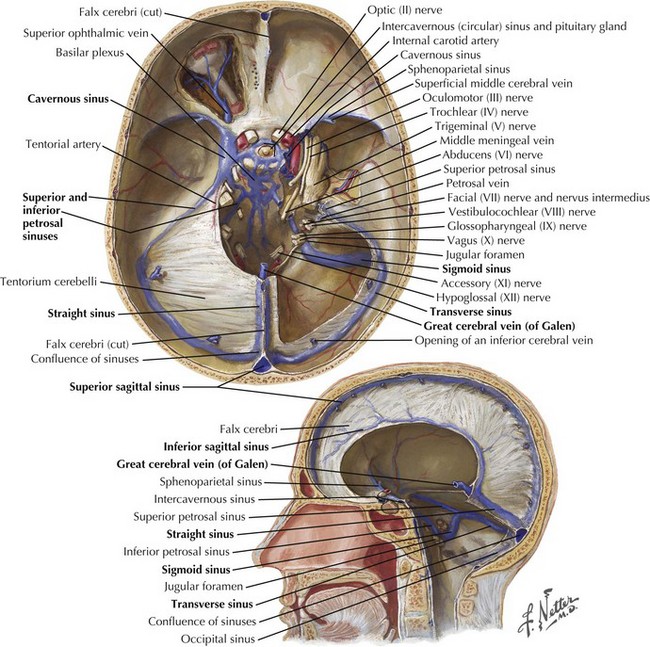
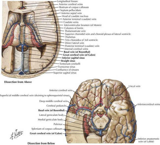
The straight sinus (Figs. 56-1 through 56-4) is formed by the splayed falx layered over the cerebellar tentorium. The inferior sagittal sinus runs in the fold of the lower arch of the falx cerebri and joins the cerebral vein of Galen in the proximity of the posterior horns of the lateral ventricles to form the straight sinus. The superior and inferior sagittal sinuses provide drainage for the cerebral hemispheres.


Stay updated, free articles. Join our Telegram channel

Jun 4, 2016 | Posted by in NEUROLOGY | Comments Off on Cerebral Venous Thrombosis

Full access? Get Clinical Tree

Get Clinical Tree app for offline access