20 Cervical and Thoracic Spine Degenerative Disease
I. Key Points
– Cervical and thoracic degenerative disease is a chronic condition but can present as acute.
– Magnetic resonance imaging (MRI) is usually the imaging modality of choice for diagnosis.
– Radicular and axial pain may be treated conservatively, but myelopathy or worsening neurologic function generally requires surgical intervention.
II. Cervical Disc Herniation
– Background
• Dehydration and fragmentation of the nucleus pulposus of cervical discs with age are natural processes.1
• The annulus and often the posterior longitudinal ligament tear, allowing the nucleus to herniate into the spinal canal, where it may compress the cord or the adjacent root at its foramen.1
• Acute disc rupture occurs more often laterally in the spinal canal due to the relative weakness of the posterior longitudinal ligament in that area; as a result, root compression occurs more often than cord compression.1
• Infarction of the cord and root may occur if compression and ischemia are severe, although it is rare.1
– Signs, symptoms, and physical exam
• Lateral disc herniations cause pain that radiates from the neck to the shoulder/arm and into the hand; the disc usually impinges on a nerve exiting from the neural foramen at the level of the herniation (e.g, a C6-C7 disc is associated with C7 radiculopathy).
• C5 symptoms: shoulder abduction (deltoid) weakness, shoulder paresthesias, deltoid and pectoralis reflexes diminished2
• C6 symptoms: forearm flexion (biceps) weakness; upper arm, thumb, and radial forearm sensory alteration; biceps and brachioradialis reflexes diminished2
• C7 symptoms: elbow extension (triceps) weakness; second and third digit sensory alteration; triceps reflex diminished2
• C8 symptoms: hand intrinsic muscle weakness; fourth and fifth digit sensory alteration; finger jerk reflex diminished2
• Central disc herniation can cause myelopathy and central cord syndrome.
– Workup
• Complete history and physical
• Basic laboratory studies
– Neuroimaging
• Based on localization of signs and symptoms
• MRI is the imaging modality of choice for visualizing soft tissue and the spinal cord/nerve roots.
• Computed tomography (CT) myelogram: when MRI cannot be done or when better bone imaging is needed
• Plain CT: good for bone imaging
• Plain x-rays: good for bone imaging, anteroposterior (AP)/lateral views useful for visualizing alignment, flexion/extension views useful to assess subluxation/instability
– Treatment
• Over 90% of patients with acute cervical radiculopathy due to cervical disc herniation can improve without surgery.2
• Surgery is indicated for those who fail to improve or those with progressive neurologic deficit who are undergoing nonsurgical management.
• Anterior surgical options: anterior cervical discectomy with or without fusion, plating, or artificial disc (arthroplasty), anterior cervical foraminotomy
• Posterior surgical options: posterior cervical laminectomy/ foraminotomy with or without fusion, keyhole laminectomy (for lateral “soft disc” herniation)
– Surgical pearls
• Partial or complete corpectomy may be required if herniated disc is sequestered posterior to vertebral body and is not accessible by discectomy.
III. Cervical Spondylotic Myelopathy
– Background
• Caused by the reduction in the sagittal diameter of the cervical spinal canal as a result of congenital and degenerative changes3
• Often due to degeneration of the intervertebral disc producing a focal stenosis due to a “cervical bar,” which is usually a combination of osteophytic spurs and/or protrusion of disc material2
• Most common type of spinal cord dysfunction in patients over the age of 55 years2
• Cord injury likely occurs as the result of several interrelated factors: direct compression of the cord, microtrauma associated with neck flexion and extension, and vascular injury.4
• Risk factors include cigarette smoking, frequent lifting, and diving.
• Signs and symptoms may overlap with those of amyotrophic lateral sclerosis (motor neuron disease).2
– Signs, symptoms, and physical exam
• Gait disturbance, often with lower extremity weakness or stiffness, is a common early finding.4
• Cervical pain and mechanical signs are uncommon in cases of pure myelopathy.
• Typical earliest motor findings are weakness in the triceps and hand intrinsic muscles.
• Clumsiness with fine motor skills (writing, buttoning buttons)4
• Glove distribution sensory loss in the hands or several levels below the area of cord compression
• Reflexes are hyperactive at a varying distance below the level of stenosis; pathologic reflexes may be present (e.g., Hoffmann, Babinski, clonus).
• Central cord syndrome, in which motor and sensory deficits affect the upper extremities more than the lower extremities, may occur acutely after trauma with hyperextension in those with cervical stenosis.
– Workup
• Complete history and physical
• Basic laboratory studies
– Neuroimaging
• Based on localization of signs and symptoms
• MRI is the imaging modality of choice for visualizing soft tissue and the spinal cord and nerve roots, but cannot distinguish between disc and bone (Fig. 20.1).
• CT myelogram: when MRI cannot be done or when better bone imaging is needed. Can still visualize spinal cord and nerve roots, but does not provide information about changes within the spinal cord parenchyma. Risks of lumbar puncture and/or intrathecal contrast injection need to be considered.
 Plain CT: good for bone imaging and may demonstrate narrow canal, but does not provide adequate information regarding soft tissues.
 Plain x-ray: good for bone imaging, may demonstrate narrow canal (posterior vertebral line to spinolaminar line <12 mm). Flexion/extension view may demonstrate dynamic instability.
– Treatment
• Nonoperative management (prolonged immobilization with rigid cervical bracing, eliminating “high-risk” activities, bed rest, and antiinflammatory medications) may be considered for mild myelopathy.
• More severe myelopathy should be treated with surgical decompression.
• Surgical approaches
 Posterior—not ideal for correction of kyphotic deformity
 Laminectomy alone (higher incidence of late kyphotic deformity)
 Laminectomy + instrumentation/fusion (lateral mass screws, etc.)
 Laminoplasty (if patient has myelopathic symptoms without axial neck pain)
 Anterior—ideal for correction of kyphotic deformities
 Anterior cervical discectomy and fusion (ACDF)
 Corpectomy and fusion: when compression extends beyond region of disc space
 Combination of ACDF + corpectomy and fusion
 Anterior procedures that include more than three disc levels will need posterior instrumentation/fusion in addition for stability.
– Surgical pearls
• Bone imaging (CT or x-ray) is important for detecting ossified posterior longitudinal ligament (OPLL) when suspected based on MRI. If present, may influence approach (posterior instead of anterior) or procedure (corpectomy instead of ACDF) to prevent intraoperative durotomy.
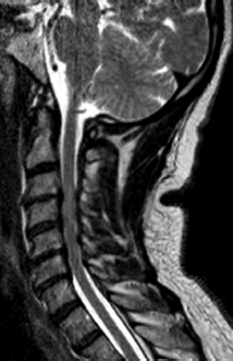
Fig. 20.1 Sagittal T2-weighted MRI of the cervical spine demonstrating multilevel degenerative disc disease, canal stenosis, and signal change in the cord.
IV. Thoracic Disc Herniation
– Background
• Incidence of clinically significant herniation is 1 patient per 1 million people.5
• 0.25% of all herniated discs2
• <4% of operations for all herniated discs5
• 75% occur below T8; most common at T11/T122
– Signs, symptoms, and physical exam
• Axial pain may be mechanical. Can sometimes be confused with cardiac, pulmonary, or abdominal pathology.
• Lateral or centrolateral herniations can present with radicular pain around chest wall along the path of an intercostal nerve in a dermatomal pattern.
• Central herniations are associated with a high incidence of spinal cord compression and long tract signs (lower extremity hyperreflexia, Romberg sign, Babinksi reflex, clonus, ataxic gait, loss of rectal tone, and decreased perianal sensation).
• In severe cases, the lesion may cause loss of bowel or bladder function and progress rapidly to incomplete or total flaccid paraplegia.
– Workup
• Complete history and physical
• Basic laboratory studies
– Neuroimaging
• Based on localization of signs and symptoms
• MRI is the imaging modality of choice for visualizing soft tissue and the spinal cord and nerve roots, but it cannot distinguish between disc and bone; scout film with MRI of entire spine needed to localize level precisely.
• CT myelogram: when MRI cannot be done, or when better bone imaging is needed along with ability to still visualize spinal cord and nerve roots; does not provide information about changes within the spinal cord parenchyma; can show calcification in disc (occurs in 30 to 70% of symptomatic thoracic discs); risks of intrathecal contrast injection need to be considered
 Plain CT: good for bone imaging but does not provide adequate information regarding soft tissues
 Plain x-ray: good for bone imaging and essential as an intraoperative reference to determine correct level
 Intraoperative fluoroscopy: it is often easier to count vertebrae upward from the sacrum or to use the ribs as a reference than to count down from C1. Use AP to localize since lateral is difficult to obtain, especially in upper/mid-thoracic spine. If doubt persists, intraoperative myelography can be performed to identify the correct level, but risks of intrathecal contrast injection need to be considered.
– Treatment
• Asymptomatic thoracic disc herniations without evidence of spinal cord compression require no treatment.
• Symptomatic thoracic disc herniations without evidence of spinal cord compression should initially be treated nonoperatively (at least 4 to 6 weeks).
 Acute herniations resulting in axial pain
 Activity modification
 Nonsteroidal antiinflammatory drugs (NSAIDs)
 Physical therapy
 Radicular pain/paresthesias
 Oral corticosteroids
 Epidural steroid injections
 Surgery may be considered for unrelenting symptoms despite nonoperative treatment.
• Symptomatic thoracic disc herniations causing spinal cord compression should be treated surgically.
 Approaches
 Anterior—good for midline or broad-based herniations or densely calcified disc herniations
 Transsternal or via resection of medial aspect of clavicle (for upper thoracic lesions)
 Anterolateral—good for midline or broad-based herniations or densely calcified disc herniations
 Transthoracic transpleural via thoracotomy (usually right-sided to avoid heart)
 Video-assisted thoracoscopic (VATS)—not widely used
 Minimally invasive transthoracic transpleural or retropleural
 Posterolateral—good for lateral or soft disc herniation
 Transpedicular
 Costotransversectomy
 Lateral extracavitary
 Posterior (laminectomy)—not recommended due to high incidence of neurologic injury
– Surgical pearls
• The anatomy involved in surgery for thoracic herniated discs is not often encountered by the spine surgeon, who must understand this anatomy thoroughly before taking a patient to the operating room.
• Calcified discs are difficult to treat via posterolateral approaches, so consider an anterior or anterolateral approach for these lesions.
• Ensure that the disc level is precisely localized by intraoperative fluoroscopy before proceeding with bone removal and discectomy.
• Posterolateral approaches may require instrumentation, especially if performed bilaterally.
• Consider somatosensory evoked potentials (SSEPs) and motor evoked potentials (MEPs) intraoperatively, especially if myelopathy is present.
Common Clinical Questions
1. A C6/C7 lateral disc herniation will compress which nerve root and cause what physical exam findings?
2. On lateral C-spine x-ray, what spinal canal diameter is considered to be stenotic?
3. Is laminectomy the best approach for excising a herniated thoracic disc?
Related posts:


Stay updated, free articles. Join our Telegram channel

Full access? Get Clinical Tree


