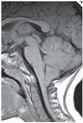
Chiari Malformation and Syringomyelia


Chiari Malformation and Syringomyelia


Rodolfo Hakim

Mark G. Luciano


The most frequently encountered forms of Chiari malformation are Chiari I and II malformations. Both are characterized by a congenital downward displacement of the cerebellum and crowding of the cervicomedullary junction. Since abnormalities of this area can disturb the entire neuroaxis via effects on CSF flow, cranial nerves, brainstem, and cerebellum, symptoms can vary widely. As a result, the identification of truly symptomatic Chiari malformation is not always straightforward. Additionally, the approach to surgical treatment has been particularly varied and controversial and has been greatly influenced by conflicting theories of pathophysiology.


CLASSIFICATION

The Chiari malformations are composed by a somewhat incongruous group of disorders of the rhombencephalon, which have as a common denominator a congenital anomaly of the cerebellum. Cleland, in 1883, published a description of patients who had hindbrain herniation, downward displacement of the cerebellar vermis, and hydromyelia; this description was consistent with a Chiari II malformation (1). In 1891, Hans Chiari, a professor of Morbid Anatomy at Charles University in Prague, described these anomalies (Chiari types I, II, and III), each with progressive increased severity of the hindbrain anomaly and associated with hydrocephalus as well (2). Chiari III was distinguished by a cystic malformation of the fourth ventricle. In 1896, he described the fourth type (Chiari type IV) in which there was no herniation of the posterior fossa contents through the foramen magnum, but instead, there was hypoplasia of the cerebellum (3). In 1907, Schwalbe and Gredig, students of Arnold, published a paper on this topic and thereby created the eponym of “Arnold-Chiari malformation” (4). Presently, these entities are more commonly known simply as a “Chiari malformation.”

It should be noted that while historically, the Chiari malformations were classified into four types using roman numerals (Chiari types I, II, III, and IV), this was never meant to be a numerical classification of severity within a single disease. In spite of this, two additional groups or types (“0” and “1.5”) have been suggested in recent years for patients in whom the severity of cerebellar herniation could not be classified precisely into either the Chiari I or Chiari II. The understanding and acceptance of these Chiari types is still controversial. They are, however, described along with the traditional categories in Table 39.1. Because Chiari III and IV are rare, this chapter focuses on Chiari I, II, their variants, and accompanying syringomyelia.


CLINICAL MANIFESTATIONS


CHIARI I

The clinical presentation and symptomatology of the Chiari I malformation is varied. The most common symptom is headache. Also, ocular, otoneurologic, brainstem, lower cranial nerve, cerebellar, and spinal cord disturbances can occur. The headache is mostly described to range from a light pressure to an intense suboccipital pounding that mostly radiates upward toward the vertex and retro-orbital region and downward toward the neck and shoulders. Characteristically, the headaches tend to worsen with Valsalva maneuvers, by lowering the position of the head and/or by sudden changes in body position, and with exercise.

The ocular symptoms that have been found in these patients are (Table 39.2) photophobia, blurred vision, diplopia, phosphenes, visual blackouts, and visual field cuts. On the fundoscopic examination, papilledema and decreased or absent venous pulsations in one or both eyes can be found; extraocular muscle palsy can also occur. The otoneurologic symptoms may include loss of equilibrium, vertigo, tinnitus, dizziness, nausea and vomiting, oscillopsia, and hypoacusia or hyperacusia. On the examination, nystagmus can also be found (lateral, downbeat, and/or rotary) (7, 8, 9, 10, 11, 12, 13, 14, 15, 16, 17, 18, 19, 20, 21, 22, 23, 24, 25, 26, 27 and 28).

The brainstem, lower cranial nerve, and cerebellar symptoms that can occur are sleep apnea, dysphagia, syncope, hoarseness/dysarthria, facial pain/numbness, throat pain, palpitations, shortness of breath, and various degrees of incoordination. On the exam, an impaired gag reflex, vocal cord paralysis, decreased trapezius muscle strength, hypertension, dysmetria, and truncal ataxia can be found.


TABLE 39.1 Brief Comparative Description of the Various Chiari Types (I-IV, “0,” and “1.5”)


Chiari “0”


Chiari Type I


– The posterior cranial fossa appears crowded, but


– Caudal displacement (herniation) of the


there is no hindbrain herniation.


cerebellar tonsils >5 mm below the foramen


– The cerebellar tonsils are within the normal range


magnum (specifically McRae’s line)


of location (no tonsillar herniation).


– The fourth ventricle and the brainstem are in


– The possible presence of a spinal cord syrinx with clinical symptoms or with an increase in its size during follow-up.


the posterior fossa. – Syringomyelia may be present (estimated at 12%) (5).


– Hydrocephalus, although uncommon, can occur probably due to an increase in the CSF outflow resistance at the level of Luschka and Magendie.


Chiari Type “1.5”


Chiari Type II


– Elements of Chiari I and II


– Caudal displacement of the brainstem,


– The cerebellar tonsils are caudally displaced >5 mm


cerebellar vermis, and fourth ventricle


below the foramen magnum (like the Chiari I type). – The brainstem and fourth ventricle are caudally displaced (like the Chiari II type). – Not associated with neural tube defects


– Associated with neural tube defects (myelomeningoceles) – Syringomyelia may be present. – (see also Table 39.2)


Chiari Type III


Chiari Type IV


– Very unusual (0.6%-4% of all the Chiari malformations). There are 42 cases reported in


– Rarely seen – Cerebellar and tentorium hypoplasia or aplasia


the literature.


– There is no hindbrain herniation.


– There is herniation of the posterior fossa contents


(cerebellum, fourth ventricle, brainstem, and occasionally the upper cervical spinal cord) through a bone defect at the level of low occipital


and or upper cervical regions.


– An encephalocele must be present in conjunction with the Chiari II brain anomalies.


– Usually associated with hydrocephalus, syringomyelia, and tethered cord (6).


The symptoms related to the spinal cord, caused by ventral spinal cord compression, dorsal cord compression, and/or a cord syrinx, are motor (spasticity, muscular weakness, and atrophy), sensory (hyperesthesia, hypoalgesia, anesthesia, burning dysesthesia, decreased positional sense, and temperature sensation), urinary and/or fecal incontinence, and impotence. On the exam, muscular weakness, muscular atrophy, decreased hand fine-motor ability, trophic phenomena dissociated sensory loss, hyperreflexia or hyporeflexia, and Babinski sign can be found. Scoliosis from syringomyelia can occur; a left thoracic curve as well as abnormal abdominal cutaneous reflexes sometimes can differentiate it from an idiopathic-type scoliosis.


CHIARI II

The Chiari II is only found and universally associated with the myelomeningocele patient population. Therefore, one can make a diagnosis of a Chiari II, even if no symptoms are present, in a newborn with a myelomeningocele. In the neonate with either Chiari I or Chiari II, the most common signs that can be found are stridor, apnea, poor feeding, and opisthotonos. In the Chiari II, within the respiratory difficulty complex, the most noticeable sign is inspiratory stridor. This is produced by a dysfunctional 10th cranial nerve and thus manifests in vocal cord paralysis.

In infants and young preschoolers, the symptomatology of the Chiari II malformation is most frequently related to brainstem compression and therefore can be a neurosurgical emergency. There can be respiratory difficulties, inspiratory stridor, apnea, aspiration pneumonia, nasal regurgitation, weight loss, prolonged feeding time, achalasia, extremity paresis, hypotonia, and opisthotonos (29, 30, 31, 32, 33 and 34).

In older children and adults, the most common symptoms are those caused by a cervical myelopathy, and therefore, one can find spasticity and upper-extremity weakness, occipitocervical pain, changes in their handwriting, and loss of muscle bulk. Also, there can be truncal and appen-dicular ataxia, dysarthria, nystagmus, difficulty in convergence, and Parinaud’s syndrome. Scoliosis, disassociated thermoalgesia, and back pain should prompt suspicion for syringomyelia.


PATHOPHYSIOLOGY

Although Chiari I and Chiari II are two different entities, they share in common the fact that there is a tight or crowded posterior fossa. This is more evident in the Chiari II since the tentorium is low and there is downward displacement of other structures besides the cerebellar tonsils. The craniocerebral disproportion creates a “pushing” or a “mass effect” and therefore alters the hydrodynamic forces
on the neural structures as well. Besides the “pushing” force, when these entities are accompanied by a tethered cord, then a “pulling” force may exist creating, as well, hydrodynamic changes at least at the level of the foramen magnum. Even though the neural structures are displaced, there are other nonneural issues that can occur and play a role in the “narrowing” of the CSF spaces surrounding the neural structures at the level of the foramen magnum. Some of the concomitant anatomical abnormalities with which the Chiari I malformation can be found associated are basilar invagination, Klippel-Feil syndrome, atlanto-occipital fusion, odontoid retroflexion (mentioned previously), Pierre-Robin syndrome, hemihypertrophy, craniofacial dysostoses, craniosynostoses, acromegaly, neurofibromatosis, mucopolysacharidoses, and Paget’s disease (12,18,35).


TABLE 39.2 Clinical Signs and Symptoms (in Decreasing Order of Frequency) Associated with Chiari Malformation


A. Suboccipital Headache: 81%


Clinical Signs:


Impaired gag reflex


B. Ocular: 78%


Facial hypoesthesia


Symptoms:


Vocal cord paralysis


Retro-ocular pain


Decreased trapezius muscle strength


Phosphenes


Glossal atrophy


Transient visual obscurations


Hypertension


Visual blackouts


Trigeminal and/or glossopharyngeal neuralgia


Blurred vision


Photophobia


E. Cerebellar


Diplopia


Symptoms:


Visual field cuts


Tremor


Clinical Signs:


Decreased fine motor function


Decreased visual acuity


Incoordination


Decreased or absent fundoscopic venous pulsations


Clinical Signs:


in one or both eyes


Dysmetria


Extraocular muscle palsy


Truncal and appendicular ataxia


Papilledema


Visual field defect


F. Spinal Cord: 94% in patients with syringomyelia; 66% in those


without


C. Otoneurologic: 74%


Symptoms:


Symptoms:


Motor:


Dizziness


Decreased strength


Loss of equilibrium


Spasticity


Tinnitus


Atrophy


Vertigo


Incontinence (urinary and/or fecal)


Nausea and vomiting


Impotence


Oscillopsia


Sensory:


Hypo- or hyperacusia


Hyperesthesia or hypoesthesia


Clinical Signs:


Anesthesia


Nystagmus (lateral, downbeat, or rotary)


Burning dysesthesia


Sensorineural hearing loss


Decreased positional sense and temperature sensation


D. Brainstem: 52%, together with Cerebellar (E.)


Clinical Signs: Decreased hand fine-motor ability


Symptoms:


Muscular weakness


Dysphagia Sleep apnea


Hyper- or hyporeflexia Babinski sign


Dysarthria


Trophic phenomena


Palpitations


Dissociated sensory loss


Throat pain


Muscular atrophy


Facial pain/numbness


Hoarseness


Syncope


Shortness of breath


Milhorat TH, Chou MW, Trinidad EM, et al. Chiari I malformation redefined: clinical and radiographic findings for 364 symptomatic patients.


Neurosurgery 1999;44:1005-1017.


One of the most important is the relation or location of the odontoid process in regard to the brainstem. When the odontoid is retroflexed, the neural structures at the level of the foramen magnum are going to have less “free CSF space” surrounding them, and also, there can be direct neural compression from the bone structures.

Syringomyelia can be found in both Chiari I and Chiari II in 12% to 75% of the cases in different series (5,36). The syrinx is most commonly found in the cervical spinal cord, but it can occur in the thoracic and lumbar spinal cord as well as in the medulla. In a study of Chiari I patients by Tubbs et al., it was found that syringomyelia, especially holocord syrinxes, was frequently associated in those patients with higher grades of odontoid retroflexion. Also, the more prominent retroflexed odontoids were more common in female patients, and also frequently, the
obices were displaced caudally greater than three standard deviations below the normal (37).


CHIARI I

The etiology of the Chiari I malformation has been suggested to be a disorder of the paraxial mesoderm in which there is an underdevelopment of the posterior cranial fossa and therefore an overcrowding of its normally developed contents (12). This has been supported by volumetric measurements of the posterior cranial fossa. As a result, the normally developed hindbrain begins to squeeze downward through the foramen magnum and therefore manifests anatomically as a tonsillar herniation (Fig. 39.1); occasionally, one can see an upward shift of the tentorium as well (38). Direct compression of the nervous structures is also evident. The restriction of the CSF to flow caudally as well as rostrally through the foramen magnum may also play a role in the production of the clinical manifestations. Also, although still controversial, it has been suggested that tonsillar herniation can occur from the constant pull or traction by a tethered spinal cord during development. Although in most cases a genetic component has not been identified, genetic components with autosomal dominant or recessive inheritance have been documented in families in which more than one member has been diagnosed with a Chiari I malformation.


Figure 39.1. Sagittal T1 MRI of an 18-month-old patient diagnosed with a Chiari I malformation showing a 9-mm tonsillar caudal displacement. The patient’s symptoms were difficulty swallowing and breathing, as well as frequent respiratory infections since early in life; worsening of the gait (unsteady with frequent falling); and during the last 2 months, posterior head and neck pain.

A secondary or acquired form of Chiari I has been found in patients with hydrocephalus, posterior fossa tumors or cysts, CNS infections, pseudotumor cerebri, spinal CSF fistulas, multiple lumbar punctures, and/or lumboperitoneal shunts (39, 40, 41 and 42). Welch et al. (42) documented in 1981 a descent of the cerebellar tonsils in patients who had undergone lumboperitoneal shunting; they therefore assumed that the cause of this anomaly was an imbalance between the intracranial and intraspinal pressures.


CHIARI II

There are four groups of theories attempting to explain the pathophysiology leading to the Chiari II malformation. The first one, postulated by Chiari, points to hydrocephalus as the cause of the hindbrain herniation (2). Although this mechanism makes sense, the literature has shown that between 10% and 20% of the patients with myelomeningocele and Chiari II do not develop hydrocephalus (35). In addition, hydrocephalus certainly does not explain other anomalies found in these patients (43).

The second theory, proposed by Cleland, is a low-volume posterior fossa due to primary hindbrain dysgenesis (1). This theory could certainly explain the herniation findings in these patients. On the other hand, it does not explain (although a theory does not necessarily need to explain all the anomalies for it to be valid) the frequently associated supratentorial and cranial anomalies found in Chiari II and myelomeningocele (42,44,45).

Only gold members can continue reading. Log In or Register to continue

Stay updated, free articles. Join our Telegram channel

Jul 5, 2016 | Posted by in NEUROSURGERY | Comments Off on Chiari Malformation and Syringomyelia

Full access? Get Clinical Tree

Get Clinical Tree app for offline access