Cranial Nerve II

4 Cranial Nerve II


Optic Nerve and Visual System


Intraocular Optic Nerve


Clinical Vignette


A 48-year-old man was referred for sudden loss of vision in the left eye. He had noted this the morning before while shaving when he could not see the lower half of his chin with the left eye only. He had no pain, and had no preceding systemic symptoms. His past medical history was noteworthy for mild diet-controlled hypercholesterolemia and untreated labile hypertension. The affected eye had 20/40 central acuity, and an inferior central field loss that extended nasally but did not cross into the superior field. The left optic nerve showed acquired elevation and swelling, with mild peripapillary hemorrhages. The fellow nerve was small in diameter, had no physiologic cup, and had mild congenital elevation. The diagnosis of idiopathic (nonarteritic) anterior ischemic optic neuropathy (AION) was made. Over the next 6 weeks, the left optic nerve swelling abated and was replaced by mild pallor noted superiorly. The vision did not recover.


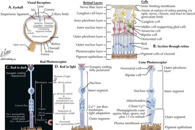
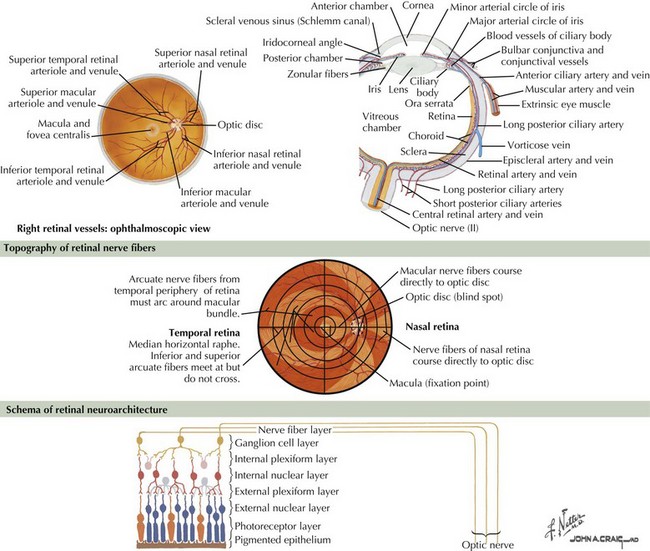
The optic nerve is not a peripheral nerve but rather a central nervous system (CNS) tract containing central myelin formed by oligodendrocytes. It is composed of long axons, whose cell bodies comprise the ganglion cell layer of the inner retina (Figs. 4-1 and 4-2). The axons run in the retina’s nerve fiber layer to gather at the optic disk.


The optic nerve nominally begins when the axons of the ganglion cells (the nerve fiber layer of the retina) turn 90°, changing orientation from horizontal along the inner retinal surface to vertical, passing through the outer retina via the scleral canal (Fig. 4-3). The gathering of axons at the canal forms the optic disk (also, optic nerve head) of the fundus. Myelin is usually absent from the nerve fiber layer where the nerve exits the globe.


Vascular supply of the retina comes from the ophthalmic artery off the internal carotid artery. Proximal branches from this artery and branches off the muscular arteries constitute the posterior ciliary arteries that form a plexus of vessels around the lamina cribrosa and supply the optic disc, the adjacent optic nerve, and the outer layers of the retina. Cilioretinal branches from this plexus often supply the macula as well. Another branch of the ophthalmic, the central retinal artery, enters the distal optic nerve and emerges out the disc dividing into four arteriolar branches to supply each quadrant of retina. The proximal part of the optic nerve is supplied by a series of small vessels of the ophthalmic while the posterior optic nerve and the chiasm have additional supply from the anterior cerebral and the anterior communicating arteries.


The shape of visual field deficits due to vascular compromise of the inner retina is predictable, being consistent with the specific location of the arterial occlusion. Visual field defects are inverted in relation to the pathologic location: for example, a superior branch occlusion of the retinal artery will cause an inferior field defect. When retinal arteriolar occlusions affect the nerve fiber layer, field defects typically extend beyond the local occlusion in an arcuate or sectoral pattern, following the arc of the nerve fiber layer. Disease of the anterior optic nerve is an important health care problem. Glaucoma alone is suspected to affect 3 million patients, accounting for 120,000 cases of blindness in the United States, with an annual governmental cost of $1.5 billion in expenditures and lost revenue.


Clinical Presentations


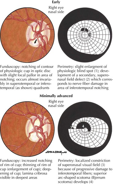
Primary open-angle glaucoma (POAG) is a chronic, progressive, degenerative disease of the optic nerve. Its usual hallmark is high intraocular pressure (IOP; above 21 mm Hg), but glaucoma without high IOP (normal pressure or low-tension glaucoma) is occasionally seen, especially in the elderly. The typical optic nerve finding is cupping atrophy (i.e., enlargement of the disk’s central cup as nerve fibers are lost), coupled by progressive visual field loss that often starts nasally, progresses superiorly and inferiorly, and finally extinguishes the central and temporal fields (Fig. 4-4). POAG is usually bilateral and asymmetric and the visual loss is permanent. The time course is measured in years, and because of the slow pace and the late involvement of the central field, patients may remain asymptomatic until the disease is quite advanced. It is essential that all standard eye examinations include screening IOP measurements and optic disk inspection.


Glaucoma has other forms besides POAG, which may be congenital, secondary to systemic disease (e.g., diabetes), or other acquired eye conditions (e.g., trauma). Among these, acute narrow-angle glaucoma (also, acute angle closure glaucoma) may present dramatically with nausea, unilateral headache, and ipsilateral monocular visual loss. The diagnosis and treatment of glaucoma forms a significant subspecialty within ophthalmology, but treatment efforts revolve around lowering of IOP, whether by medical or surgical means. There are no restorative or neuroprotective treatments.


Central retinal artery occlusion (CRAO) results from interruption of the central retinal artery circulation with ischemia to the entire retina. If only a portion of the inner retinal circulation is affected, a more limited version, branch retinal artery occlusion (BRAO), is present. BRAO and CRAO are in effect retinal strokes, affecting the nerve fiber and ganglion cell layers. The presentation is one of sudden, painless, complete or partial monocular visual loss often described as a “curtain” obscuring the involved area. Retinal infarcts are commonly caused by emboli, and in BRAO the embolus is typically visible in the affected retinal vessel. Episodes of temporary monocular visual loss (TMVL; transient monocular blindness or TMB and also, amaurosis fugax) often herald retinal infarcts and represent temporarily compromised flow of the inner retinal arteries usually by passing clot.


Patients who present for care within the first hours after the onset of CRAO or large BRAO are usually treated with intermittent ocular massage and lowering of IOP (either by topical agents or by paracentesis of the anterior chamber) to promote movement of the embolus to a more distal arteriolar branch. Oxygen, alone or in combination with 5% CO2 to promote arteriodilation, can also be used. Based on animal studies, it is felt that such interventions are unlikely to be helpful after 100 minutes of retinal ischemia, and in general the outlook for recovery is bleak; nevertheless, significant recovery of vision, even beyond the 100-minute window, is occasionally seen.


CRAO, BRAO, and TMVL may also serve as a warning sign of impending hemispheric stroke. Identification and treatment of the embolic source, if one can be identified, becomes the main focus of therapy after the window for acute treatment of the involved eye has passed. CRAO is often a sign of carotid stenosis, the appropriate management of which will significantly reduce long-term stroke risk (see Chapter 55, “Ischemic Stroke”). Heart embolism is another cause and a full stroke investigation is usually required. Nevertheless, up to 40% of cases remain without a definite identifiable cause with the presumed mechanism relating to intrinsic narrowing of the retinal artery due to atherosclerosis or, less commonly, other arteritides or compression.


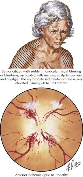
Anterior ischemic optic neuropathy can be divided into nonarteritic and arteritic (associated with temporal arteritis [TA]) and is caused by loss of blood flow in the short posterior ciliary arteries. Patients usually experience sudden and severe painless monocular visual loss, often on awakening. Examination classically reveals an altitudinal (superior or inferior) visual field loss, with a unilaterally swollen, hemorrhagic disk (Fig. 4-5). The disk loses its swelling and becomes pale within weeks. The visual loss in most cases does not change following the event but 20% may show measurable change for better or worse over days. In contrast to retinal artery occlusions, embolic AION is extremely rare. In most cases, AION occurs in middle-aged individuals who have a congenitally small, elevated (“crowded”) optic disk, or in those with one or more vascular disease risk factors, such as diabetes, hypertension, or sleep apnea. In these cases, a transient fall in blood pressure causes hypoperfusion of the posterior ciliary circulation and subsequent ischemic damage to the optic nerve head.


There is no proven treatment for AION, although a recent retrospective study suggests that acute treatment with oral steroids may improve outcome. There is a 50% risk of eventual involvement of the fellow eye. Strategies to reduce this risk have focused on identifying and treating cerebrovascular risk factors, daily aspirin, preventing systemic hypotension, and avoiding certain drugs, such as sildenafil, which may be associated with higher risk.


In older patients, AION can be a complication, and sometimes the presenting sign, of TA (also, giant cell arteritis), a systemic inflammatory process of the medium-sized arteries. TA can also produce TMVL and CRAO. Funduscopic appearance in arteritic AION often consists of pallid swelling of the disk, in contrast to the hyperemic swelling seen in idiopathic AION. In addition to an altitudinal visual loss, patients will have arteritic symptoms, including headache, scalp tenderness, jaw claudication, neck pain, malaise, loss of appetite, fevers, and morning stiffness of proximal muscles (i.e., polymyalgia rheumatica). Only rarely will a patient with arteritic AION have little or no systemic symptoms.


Untreated, TA may lead rapidly to blindness from bilateral AION, or to other serious complications including aortic dissection, myocardial infarction, renal disease, and stroke. Therefore, in any patient older than age 50 years with AION, clinical suspicion for TA is raised especially in the presence of systemic symptoms, or physical exam findings (pallid disk swelling or abnormal greater superficial temporal arteries). A high erythrocyte sedimentation rate (ESR, >45 mm/hour), high C-reactive protein (CRP, >2.45 mg/L), normocytic anemia, and thrombocytosis are supportive, but the diagnosis is established by temporal artery biopsy that reveals inflammation in the media of the arteries with disruption of the internal elastic membrane. The presence of characteristic multinucleated giant cells within the affected areas is diagnostic.


TA is treated with high-dose corticosteroids, started urgently and usually tapered over many months. Other anti-inflammatory medications, especially methotrexate, have been used in those at high risk for corticosteroid complications, but the efficacy of nonsteroidal agents has been questioned. Steroid dosage is gradually reduced over time, with the patient monitored for disease recrudescence by following symptoms and the ESR or CRP.


Papilledema (see Fig. 1-6) is bilateral optic nerve elevation and expansion due to high intracranial pressure (ICP). In mild cases, patients may have no visual symptoms. Moderate papilledema is typically accompanied by transient binocular visual obscurations, either spontaneously or during coughing, straining, or abrupt postural change. Other symptoms of high ICP may be present and include headaches (worse with recumbency) and diplopia (resulting from nonlocalizing abducens palsy; see Chapter 5). When visual loss occurs, it starts with blind spot enlargement (see Fig. 1-6), a nonspecific and often reversible change. Visual field loss resembling that of glaucoma can ensue, often over a period of many weeks. However, papilledema due to very high ICP can progress rapidly, with severe permanent visual loss within days.


Many pathophysiological mechanisms are associated with papilledema, including CNS tumor with mass effect or edema, obstructive hydrocephalus, meningitis, certain medications (e.g., tetracycline or vitamin A), and intracranial venous thrombosis or obstruction. Papilledema is occasionally seen without explanation in obese women of childbearing age and is then termed idiopathic intracranial hypertension (IIH; also, pseudotumor cerebri; see Chapter 11). Treatment involves only weight loss if the condition is mild and there is no evidence of progressive visual loss or debilitating headache. In progressive IIH, in addition to weight loss, carbonic anhydrase inhibitors such as acetazolamide (typically 1–2 g/day in divided doses) are used to reduce cerebrospinal fluid (CSF) production and optic nerve edema. When medical treatment fails, two surgical options exist: optic nerve sheath fenestration or CSF shunting either with lumboperitoneal or ventriculoperitoneal shunts.


Papilledema can be mimicked by the rare entity of optic perineuritis, which consists of monocular or bilateral optic disk swelling without central visual loss or raised ICP. Its usual cause is idiopathic optic nerve sheath swelling or inflammatory orbital pseudotumor but may be due to a systemic arteritis (Wegener or giant cell arteritis) or of an infectious (syphilitic) etiology.


Optic nerve drusen are small, translucent, usually bilateral concretions within the substance of the disk that may be observed in perhaps 1% of patients. Drusen contain calcium and can therefore be demonstrated on ultrasound and computed tomographic (CT) examinations. It is speculated that a very small scleral canal may inhibit proper axonal metabolism, causing extracellular debris to be deposited as drusen over time. Drusen of the optic nerve is often associated with visual field loss, and treatment to retard such loss is uncertain. Drusen of the nerve head are occasionally seen in patients with certain retinal disorders, such as retinitis pigmentosa.


Congenital dysplasia of the optic nerve can be seen as an isolated monocular or binocular finding, or as part of a larger disorder. The mildest form of dysplasia is “tilted” optic disks: nerve heads that are overall small with the nasal portions appearing elevated; superior temporal visual field loss (sometimes mimicking bitemporal hemianopia) is often encountered. Septo-optic dysplasia combines optic nerve hypoplasia with dysgenesis of midline brain structures, often with pituitary dysfunction. Up to a quarter of patients with fetal alcohol syndrome will have disk hypoplasia with associated inferior visual field loss, among other ocular manifestations. Optic nerve coloboma (congenital incomplete or malfusion of the globe structures including the retina and optic nerve) can be part of Aicardi syndrome, and the “morning glory” disk anomaly has been associated with several developmental syndromes.


Orbital and Intracanalicular Optic Nerve


Clinical Vignette


A 26-year-old woman presented with right monocular visual loss and headache after a car accident. She said she had suffered “whip-lash,” without bruising impact to the head. The visual loss had started 2 days after the accident. The headache was centered at the right orbit, with eye movement among its aggravating factors. Subjective visual acuity was 20/80 right eye, and visual field testing revealed nonphysiologic responses, indicating the patient was inattentive to the test, in both eyes. Fundus examination of both eyes was entirely normal; however, pupillary examination suggested a mild relative afferent papillary defect on the right. A magnetic resonance imaging (MRI) examination was obtained revealing multiple white-matter lesions. A diagnosis of multiple sclerosis (MS) presenting as optic neuritis was eventually confirmed based on spinal fluid assays and subsequent clinical course.


After leaving the eye, the fibers of the optic nerve become myelinated. The optic nerve sheath invests the nerve, starting at the sclera and becoming contiguous with the intracranial dura. CSF is present within the sheath. The optic nerve lies in the central orbit within the extraocular muscle cone and exits the orbit through the optic canal before traveling a short distance intracranially to join the chiasm. Vascular supply is via branches of the ophthalmic artery.


Diseases that affect the orbital optic nerve give characteristic central visual field loss. It is believed that the nerve fibers corresponding to central vision, among the most metabolically active cells in the visual system, occupy a central position in the optic nerve, farthest away from the exterior blood supply. The central fibers, therefore, are the most prone to dysfunction or injury due to varying mechanisms, including compression, ischemia, metabolic disease, and toxic insult. Within the bony optic canal, the optic nerve is confined in a small space and is relatively immobile, making it susceptible to quite small tumors or inflammatory processes as well as shear injury produced by deceleration head trauma.


Multiple sclerosis (see Chapter 46), however, remains the chief cause of orbital optic nerve disease and is the initial manifestation in approximately 20% of patients. An additional 20% will eventually experience it throughout the course of the disease. It is estimated that more than 90% of patients suffering “isolated” optic neuritis will eventually receive a diagnosis of MS. Diagnostic testing in optic neuritis naturally mirrors that for MS, with brain MRI and CSF analysis being the primary tools.


Clinical Presentations


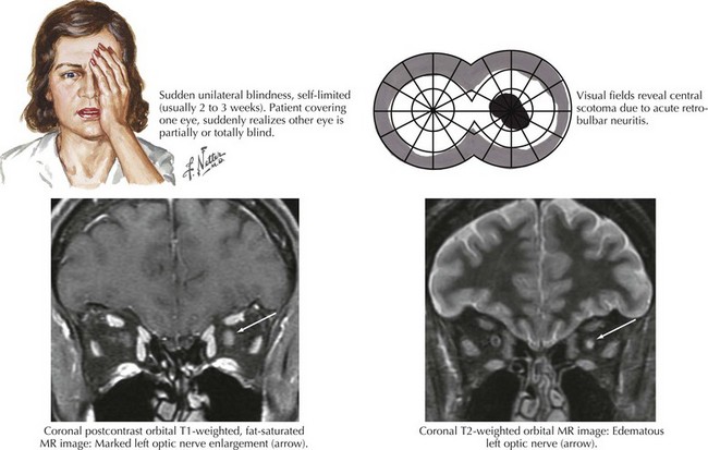
Optic neuritis is the clinical syndrome of subacute painful, monocular visual loss. The pain often precedes visual loss by a day or more and is a periorbital ache made worse with eye movements. Ensuing visual loss is often sudden and severe, with perceived worsening over several days. The degree of visual field loss varies, but a central scotoma is the classic finding (Fig. 4-6). Examination may also demonstrate loss of central acuity, contrast sensitivity, and color perception in the affected eye.


Initially, funduscopic appearance of the affected disk is normal, but only the presence of a relative afferent pupillary defect and visual loss confirms that optic neuropathy is present. Occasionally, mild ipsilateral disk swelling is seen, and in all cases some degree of optic pallor, usually localized to the temporal quadrant of the disk, appears within weeks. Incomplete recovery of vision, mostly in the first 3 months, is expected with central acuity recovering better than other parameters, often to near normal.


As with other manifestations of MS, emphasis is on early diagnosis so that patients may begin treatment with immunomodulating medications to reduce disease activity and associated morbidity. Intravenous methylprednisolone (1 g/day for 3 days, followed by an oral prednisone taper for 11 days) has been shown to accelerate visual recovery in optic neuritis, although the final level of recovery is unaffected. The same study showed a reduced risk of MS exacerbations for 2 years following methylprednisolone pulse treatment. It is unclear if the drug provides additional protection beyond 2 years and whether it affects outcome in the long run. Oral prednisone alone is contraindicated in typical demyelinating optic neuritis.


Optic neuritis can also be seen as part of Devic neuromyelitis optica, an MS-like disease defined by episodes of optic neuritis and transverse myelitis. The immunopathogenesis appears distinct from MS and the preferred therapies are parenteral corticosteroids and plasmapheresis acutely, with long-term immunosuppressive agents, such as azathioprine, used to prevent relapses. The presence of a hallmark serum immunoglobulin (NMO-IgG directed against the aquaporin-4 protein) is central to diagnosis.


Optic neuritis can occasionally be idiopathic, with prolonged surveillance never leading to a diagnosis of MS. In rare cases, optic neuritis can be mimicked by treponemal infection, or by inflammatory disease (e.g., sarcoidosis).


Posterior ischemic optic neuropathy presents as sudden, painless monocular visual loss without acute change in the ocular fundus and disk. Over weeks, disk pallor becomes evident. Classically seen in chronically anemic patients after major gastrointestinal hemorrhage, it has been more recently found in one of three clinical settings: as bilateral visual loss after major surgery; and as unilateral visual loss, either as a complication of TA or of peripheral vascular disease. There is no definitive test for posterior ischemic optic neuropathy, and diagnostic workup is directed toward ruling out arteritis and occlusive carotid disease.


Indirect traumatic optic neuropathy

Stay updated, free articles. Join our Telegram channel

Jun 4, 2016 | Posted by in NEUROLOGY | Comments Off on Cranial Nerve II

Full access? Get Clinical Tree
Get Clinical Tree app for offline access