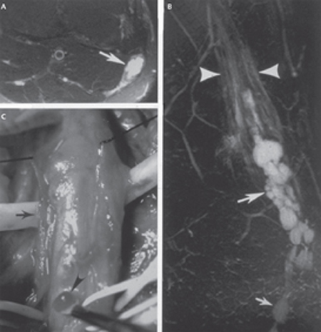
43 Diagnosis and Treatment of Common Peroneal Nerve Problems A 20-year-old man suffered a knife laceration to the lateral part of his right lower leg just below the fibular head. He immediately developed a complete right foot drop but did not see a physician until 2 weeks after the injury. His neurological exam 2 weeks after the injury revealed absent sensory and motor function in a distal right common peroneal nerve (CPN) distribution. Specifically, he could neither evert (0/5: superficial peroneal nerve [SPN] branch) nor dorsiflex (0/5: deep peroneal nerve [DPN] branch) his right foot. He also had no sensation along the dorsum of his right foot. The wound was tender and there was a positive Tinel response just proximal to the injury with dysesthesias radiating into the dorsum of his right foot and toes. Muscle strength and sensation in his right lower extremity supplied by the distal tibial nerve were normal and the ankle reflex was normal (2/4). Sensation along his right lateral malleolus in a sural nerve distribution was also normal. Magnetic resonance imaging (MRI) of the right lower leg performed 2 weeks after the laceration revealed discontinuity of the right CPN at the level of the skin laceration. There was also increased signal on short inversion time inversion-recovery (STIR) and T2 with fat suppression pulse sequences in the nerve and its branches distal to the transection injury. Two weeks after the CPN injury, electromyographic (EMG) and nerve conduction velocity (NCV) studies were done. The EMG study demonstrated 4+ spontaneous fibrillations with positive sharp waves in all lower leg muscles supplied by the right CPN distal to the short head of the biceps femoris muscle. There were no voluntary units in these muscles and no peroneal nerve conduction response distal to the injury. The NCV study indicated absence of conduction in the CPN across the laceration. The short head of the biceps femoris muscle, the only hamstring muscle supplied by the peroneal nerve, was normal indicating that the laceration of the nerve was more distal in location. A 40-year-old basketball coach noticed that his right foot was weak and would “slap” the ground when walking or running. He had pain in the anterolateral aspect of his right lower leg and the dorsum of his foot. He also experienced tingling sensations along the dorsum of his right foot. These symptoms and findings were all in the distribution of the CPN. On physical examination, he demonstrated mild weakness in everting his right foot (4/5: SPN branch) and more severe weakness in dorsiflexing his right foot and toes (2/5: DPN branch). He had normal plantar-flexion and inversion of the foot, as well as flexion of the toes. Sensation was reduced along the dorsum of his right foot, especially along the dorsal web space of his first (big) and second toes. His ankle reflexes were normal bilaterally. He also had a palpable lump with a Tinel response adjacent to the right fibular head with dysesthesias radiating into his big toe. Ultrasound examination showed a hypoechoic zone within the right CPN. The MRI evaluation across the right knee visualized multiple well-demarcated cysts within the CPN extending into the DPN. The lesions were hypointense on T1-weighted images and hyperintense on T2-weighted MRI. Some atrophy and increased signal were also apparent on T2-weighted and STIR images within anterior compartment muscles of the lower leg, suggesting selective denervation within the anterior compartment muscles of the leg supplied by the DPN [tibialis anterior (TA), extensor digitorum longus (EDL), extensor hallucis longus (EHL)]. Axial MRI slices at the level of the tibiofibular joint revealed cysts extending into an articular branch (“tail sign” of Spinner et al). An EMG study revealed muscle denervation changes that were most severe in the TA, EDL, and EHL (supplied by the DPN), and less severe in the peroneus longus (PL) and peroneus brevis (PB) (supplied by the SPN). Nerve conduction studies showed a slowing of responses in the right CPN across the fibular head. Patient #1: Common peroneal nerve injury from open trauma Patient #2: Intraneural ganglion cyst of the common peroneal nerve The CPN is the smaller lateral component of the sciatic nerve and is derived primarily from the dorsal branches of the L4, L5, S1, and S2 spinal nerves. Its most proximal branch in the thigh supplies the short head of the biceps femoris muscle. As it reaches the popliteal fossa, the CPN then crosses over the lateral head of the gastrocnemius muscle to run superficially just posterior to the head of the fibula where it winds around the neck of the fibula. It then gives off articular branches to the tibiofibular joint space and contributes a sensory branch, the lateral sural cutaneous nerve, which joins a branch from the tibial nerve, the medial sural cutaneous nerve, to form the sural nerve. The three articular branches accompany the superior and inferior lateral genicular arteries of the knee and the anterior tibial recurrent arteries. Continuing distally, the CPN then dives deep between the two heads of the peroneus longus muscle where it divides into SPN and DPN branches. The SPN supplies the PL and PB muscles in the lateral compartment of the leg, which evert the foot. It also supplies sensation along most of the dorsum of the foot except for a small dorsal region at the base of the big and second toes, which is supplied by a sensory branch from the DPN branch. The DPN supplies the TA, EDL, EHL, and peroneus tertius (PT) muscles and is responsible for dorsiflexion of the foot. During exploratory surgery, the tendon of the short head of the biceps femoris can be very useful in localizing the CPN. However, care should be taken to avoid confusing this tendon with the nerve. Injury to the CPN will cause clinical symptoms and findings of pain, dysesthesias, and loss of sensory and motor function in the lower leg. Weakness or loss of the ability to dorsiflex (foot drop) or evert the foot is a typical sign of this nerve injury. The CPN may be injured from direct compression and/or stretch (e.g., total knee arthroplasty), laceration (as in patient #1), or direct or indirect involvement by masses such as tumors (e.g., schwannoma or neurofibroma) or a ganglion cyst (intraneural as in patient #2, or extraneural). In the lower extremity, the most common site for a ganglion cyst is the peroneal nerve at the level of the knee and proximal tibiofibular joint. Systemic disorders such as diabetes mellitus or vasculitis, and hereditary diseases involving nerves, such as hereditary nerve pressure palsy, can also predispose this nerve to injury. Both patients presented with weakness in dorsiflexing and everting their foot. These findings could be the result of proximal spinal pathology (such as an L5 radiculopathy) or selective pathology within the lumbosacral plexus of the pelvis. In both cases, the tibial component of the sciatic nerve was spared, making these proximal locations less likely to be the site of pathology. The key muscle in the clinical examination when trying to distinguish CPN neuropathy versus L5 radiculopathy is the tibial-innervated posterior tibialis—if foot inversion is preserved, this implicates the CPN. Weakness of foot inversion indicates that the site of injury is more proximal (i.e., either the sciatic nerve or the L5 root). In addition, because the biceps femoris muscle was not involved, the pathology in both cases must be distal to it, and therefore involves the CPN and/or its distal branches. MRI and EMG studies were used to help confirm the diagnosis and localize as well as visualize the pathology. High-resolution MRI techniques applied to peripheral nerves subjected to trauma can show abnormally increased signal changes on certain pulse sequences (e.g., STIR and T2 with fat suppression) at and distal to the point of injury in the setting of axonal degeneration. In addition, high-resolution MRI can sometimes show whether a nerve is in continuity, and whether its fascicular structure is preserved, particularly on T1 pulse sequences. For patient #1, MRI showed the location where the CPN had been lacerated. In patient #2, MRI visualized the location and type of mass involving the CPN. Because the injury is an open laceration, the major differential diagnosis involves identifying the injured nerve (CPN) and determining the grade of injury (neurapraxic, axonotmetic, or neurotmetic in which myelin; myelin and axon; or myelin, axon, and the connective tissue highway are disrupted, respectively). Patients with masses involving the CPN also have symptoms and findings in the distribution of this nerve that can vary widely. MRI, ultrasound, and CT imaging may be used to visualize and distinguish different types of masses (e.g., cysts and tumors). MRI and ultrasound are particularly useful in identifying both extraneural and intraneural cystic structures. In the case of an intraneural ganglion cyst involving the CPN, initial symptoms often consist of poorly localized pain around the lateral aspect of the knee and the lower leg, as well as a partial motor deficit in muscles supplied by the DPN, which produces a partial foot drop. In more severe cases, the SPN portion of the CPN may become involved. In our patient, MRI and EMG studies helped locate and determine the grade of nerve injury (Fig. 43–1). The fact that the mass had a cystic appearance on MRI with no contrast enhancement identified it as a ganglion cyst. The MRI also clearly showed that the cysts had a tubular configuration and extended within the DPN and CPN. In addition, a tail could be seen, indicating its origin from the tibiofibular joint via an articular branch, further confirming the diagnosis of intraneural ganglion cyst(s).
 Case Presentation
Case 1
Case 2
 Diagnosis
 Anatomy
 Characteristic Clinical Presentation
 Differential Diagnosis
Case 1
Case 2

Stay updated, free articles. Join our Telegram channel

Full access? Get Clinical Tree


