Electrical events

7 Electrical events


Structure of the Plasma Membrane


In common with cells elsewhere, the plasma membrane of neurons is a double layer (bilayer) of phospholipids made up of phosphate heads facing the aqueous media of the extracellular and intracellular spaces, and paired lipid tails forming a fatty membrane in between (Figure 7.1). The phosphate layer is water-soluble (hydrophilic, or polar) and the double lipid layer is water-insoluble (hydrophobic, or non-polar).
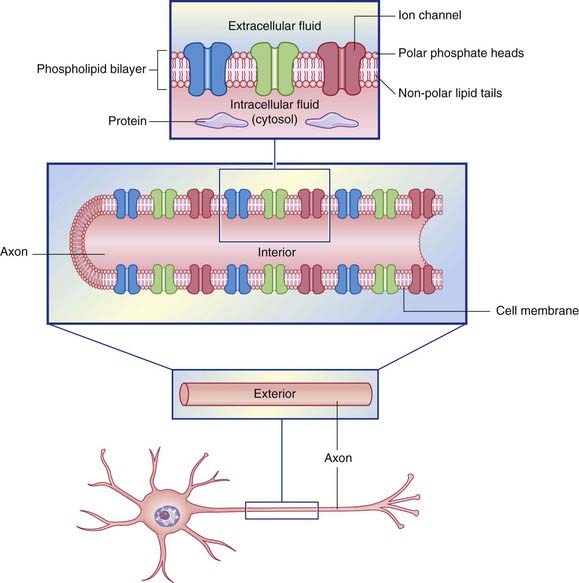


Both the extracellular and the intracellular fluids are aqueous salt solutions in which many soluble molecules dissociate into positively or negatively charged atoms or groups of atoms called ions. Ions and molecules in aqueous solutions are in a constant state of agitation, being subject to diffusion, whereby they tend to move from areas of higher concentration to areas of lower concentration. In addition to passing down their concentration gradients by diffusion, ions are influenced by electrical gradients. Positively charged ions including Na+ and K+ are called cations because, in an electrical field, they migrate to the cathode. Negatively charged ions including Cl are called anions because these migrate to the anode. Like charges (e.g. Na+ and K+) repel one another, unlike charges (e.g. Na+ and Cl) attract one another.


The cell membrane can be regarded as an electric capacitor, because it comprises outer and inner layers carrying ionic charges of opposite kind, with a (fatty) insulator in between. Away from the membrane, the voltage in the tissue fluid is brought to zero (0 mV) by the neutralizing effect of chloride anions on sodium and other cations, and the voltage in the cytosol away from the membrane is brought to zero by the neutralizing effect of anionic proteins on K+ cations.


Ion channels


Ion channels are membrane-spanning proteins having a central pore that permits passage of ions across the cell membrane. Most channels are selective for a particular ion, for example Na+, or K+ or Cl


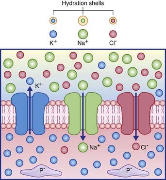
Several channel categories are recognized, of which the first three are of immediate relevance.


Figure 7.2 depicts the three passive channels concerned with generating the resting potential.


The existence of distinct channels for Na+, K+ and Cl ions would result in zero voltage difference across the membrane if passive diffusion of the three ions were equally free. However, the number of sodium channels is relatively small, and movement of the Na+ ion is relatively slow because of its relatively large ‘hydration shell’ of H2O molecules. In effect, the membrane is many times more permeable to K+ and Cl than to Na+.


The resting membrane potential


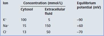
The membrane potential of the resting (inactive) neuron is generated primarily by differences in concentration of the sodium (Na+) and potassium (K+) ions dissolved in the aqueous environments of extracellular fluid (ECF) and cytosol. In Table 7.1, it can be seen that potassium is 20 times more concentrated in the cytosol; sodium is 10 and chloride 3.8 times more concentrated in the ECF.


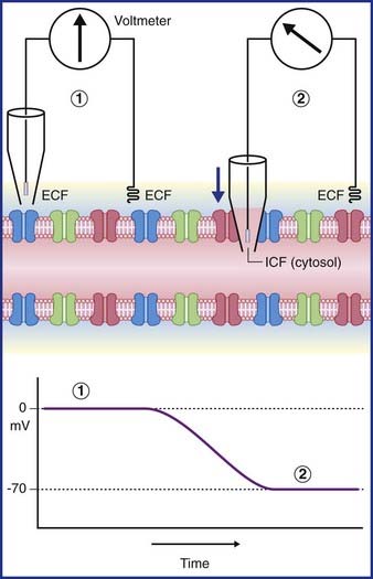
In Figure 7.3, a voltmeter is connected to electrodes inserted into the ECF surrounding an axon. One of the electrodes has been inserted into a glass pipette having a minute tip. On the left side of the figure, both electrode tips are in the ECF, and there is no voltage difference; a zero value is recorded. On the right side, the pipette has been lowered, puncturing the plasma membrane of the axon and admitting the intracellular fluid of the cytosol. The electrical charge now reveals a potential (voltage) difference of −70 mV. In practice, the membrane potential ranges from −60 mV to −80 mV in different neurons. These values represent the resting membrane potential, i.e. when impulses are not being conducted.


Jun 10, 2016 | Posted by in NEUROLOGY | Comments Off on Electrical events

Full access? Get Clinical Tree

Get Clinical Tree app for offline access