38 In the WHO classification (2007), listed embryonal neuroepithelial neoplasms are: Many embryonal neoplasms are circumscribed, pink or gray neoplasms, which may contain areas of hemorrhage, necrosis, or calcification (Fig. 38.1). CNS neuroblastomas and medulloepitheliomas sometimes contain cysts. All embryonal neoplasms have the capacity to invade the brain and spinal cord, and this will often be evident microscopically, if not macroscopically. However, infiltration occurs to a variable degree, and is rarely as diffuse as demonstrated by some astrocytic tumors. 38.1 Medulloblastoma. The texture of embryonal neoplasms varies. Some are soft, but some medulloblastomas in the lateral cerebellar cortex and some cerebral neuroblastomas tend to be firm because they contain areas of desmoplasia. Neoplastic cells occasionally metastasize through the CSF pathways (Fig. 38.2). 38.2 Supratentorial PNET in the subarachnoid space. Nevoid basal cell carcinoma syndrome (Gorlin syndrome) • Chromosome 17 abnormalities (~60%) • Isodicentric chromosome 17q (~35%) • Allelic losses on 10q (~10%) The classic medulloblastoma is composed of isomorphic cells with a high nuclear:cytoplasmic ratio (Fig. 38.3). Sheets of hyperchromatic round or oval nuclei set against a neuropil-like matrix give a monotonous appearance, with scattered mitotic figures and apoptotic bodies in the background. Necrosis is variably present, but angiogenesis with endothelial hyperplasia is a rare feature in these neoplasms. Though frequently forming a mass in the fourth ventricle, the medulloblastoma is an invasive neoplasm. Its cells have a tendency to spread along the pial surface of the cerebellum, invading the underlying cortex in swathes. Infiltration of the leptomeninges can produce a striking desmoplasia (Fig. 38.3). 38.3 Medulloblastoma.
Embryonal neuroepithelial neoplasms of the CNS
EMBRYONAL NEUROEPITHELIAL NEOPLASMS
 a tendency to disseminate through CSF pathways
 a dominant population of small undifferentiated cells
 a high mitotic index and widespread apoptosis
 Medulloblastoma and its variants:
 CNS primitive neuroectodermal tumor (PNET) and its variants:
 EMBRYONAL NEUROEPITHELIAL NEOPLASMS
 Represents about 5% of intracranial primary neoplasms.
 Represents about 20% of childhood CNS neoplasms.
 Represents over 90% of childhood CNS PNETs.
 Can present at any age, but 60% occur in patients less than 10 years of age.
 Generally midline, filling the fourth ventricle.
 Belongs to the SHH molecular subgroup, when in the lateral cerebellar cortex.
 Regarded as high-risk if a large cell or anaplastic pathological variant.
 Has a 5-year survival of 80% in standard-risk patients.
 Has a 5-year survival of 25% in high-risk patients.
 Therapies are associated with significant adverse effects, e.g. cognitive impairment.
MACROSCOPIC APPEARANCES
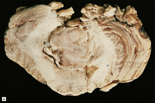
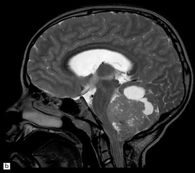
(a) A soft homogeneous mass destroys and occupies the fourth ventricle. (b) A sagittal midline MR image through the brain showing a medulloblastoma in the posterior fossa between the cerebellum and brain stem. (Dr N Sabin, St Jude Children’s Research Hospital.)
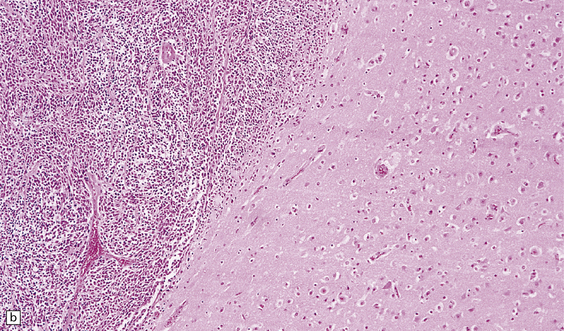
(a) There is an extensive infiltration of the subarachnoid space by basophilic cells from a PNET. Several distinct parenchymal deposits are also present. (b) From the same case, a mass of small cells fills the subarachnoid space and has begun to invade the pial surface of the cerebrum.
 FAMILIAL CANCER SYNDROMES FEATURING MEDULLOBLASTOMAS
 GENETIC ASPECTS OF MEDULLOBLASTOMA
 Common chromosomal abnormalities include:
 Gene-specific abnormalities include:
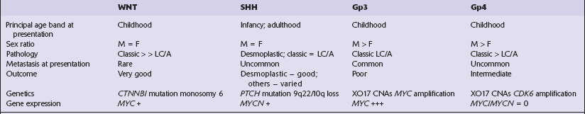
 Molecular subgroups derived from gene expression profiling have been classified into four main groups (see Table 38.1):
 Molecular favorable-outcome indicators:
 Molecular poor-outcome indicators:
MICROSCOPIC APPEARANCES
Medulloblastoma
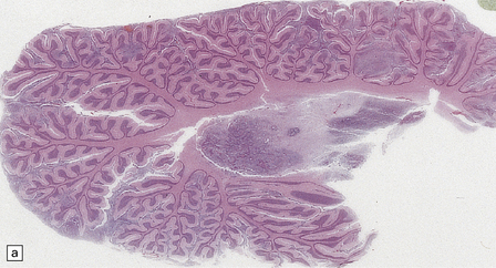
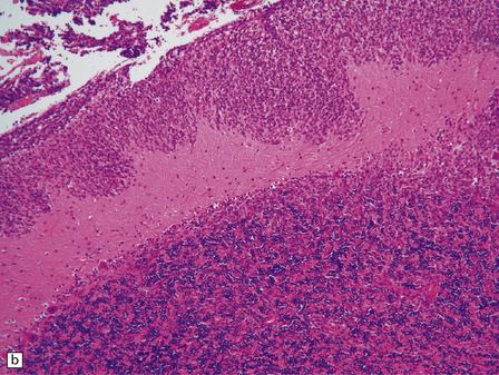
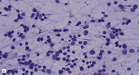
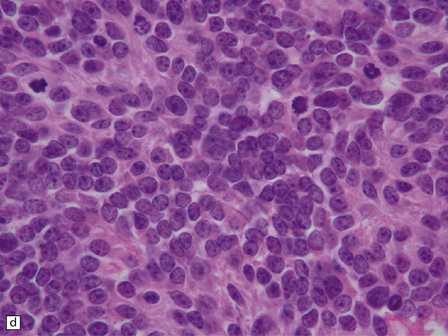
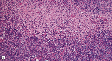
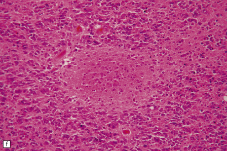
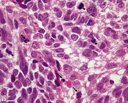
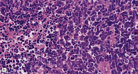
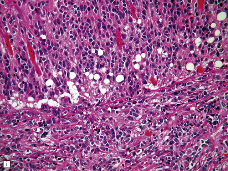
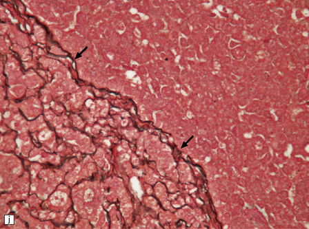
(a) Multiple tumor deposits and leptomeningeal spread of medulloblastoma cells are evident in this section of cerebellum. (b) Centripetal spread through the cerebellar cortex is a frequent finding when there is leptomeningeal disease. (c) This smear preparation shows scattered bare hyperchromatic nuclei. (d) Uniform small cells with round nuclei and a high density characterize the classic medulloblastoma. (e) A monotonous sheet of densely packed small cells is interrupted by an area with a reduced cell density. (f) Approximately 7% of classic medulloblastomas contain hypocellular nodules that contain neurocytic cells with a low growth fraction, but critically there is no internodular desmoplasia. (g) Abundant mitoses (arrowheads) and apoptotic bodies (arrows) are typical. (h) Micronecrosis. (i) Desmoplasia results when medulloblastoma cells spill out into the leptomeninges (bottom half of image). (j) The leptomeningeal desmoplasia (arrows) is demonstrated well by a reticulin preparation.![]()
Stay updated, free articles. Join our Telegram channel

Full access? Get Clinical Tree


Embryonal neuroepithelial neoplasms of the CNS


Only gold members can continue reading. Log In or Register to continue


