Enlarged Cortical Veins
James D. Eastwood, MD
DIFFERENTIAL DIAGNOSIS
Common
Normal
Developmental Venous Anomaly
Arteriovenous Malformation
Dural A-V Fistula
Less Common
Thrombosis, Dural Sinus
Thrombosis, Cortical Venous
Rare but Important
Venous Varix
Vein of Galen Malformation
Sinus Pericranii
ESSENTIAL INFORMATION
Key Differential Diagnosis Issues
Is there one enlarged vein or several?
Are they definitely veins? Could some be arteries?
Is there evidence of thrombosis?
Helpful Clues for Common Diagnoses
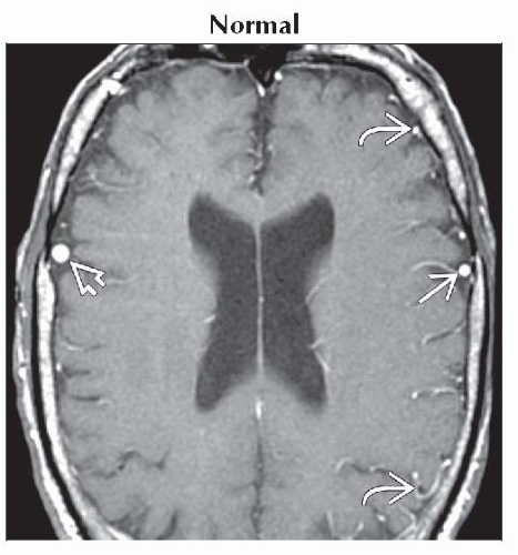
Normal
Major anastomotic cortical veins (Trolard, Labbe, SMCV) can be very prominent
Developmental Venous Anomaly
Central vein of DVA can drain cortically
“Medusa head” configuration
Arteriovenous Malformation
Prominent cortical veins tend to be regional, geographically related to nidus
Look for varices, stenoses, stagnant flow
Caution: Prominent veins may persist even if no residual AV shunting present
Dural A-V Fistula
Chronic dural sinus thrombosis (vascularized thrombus) common precursor
Cognard type IIB shows reflux into cortical veins; types III-IV have direct cortical drainage
Hemorrhage risk ↑
Helpful Clues for Less Common Diagnoses
Thrombosis, Dural Sinus
Thrombosis/stenosis causes increased back pressure
T2* (clot “blooms”), CECT or T1 C+ MR (“empty delta” sign) helpful
Thrombosis, Cortical Venous
Can occur without dural sinus occlusion
Hyperdense on NECT (“cord sign”)
T2* most useful (“blooms”)
Helpful Clues for Rare Diagnoses
Venous Varix
Usually with AVM or dAVF
Isolated venous varices are rare
Vein of Galen Malformation
Deep veins but cortical may ↑ if large
Sinus Pericranii
Transcalvarial communication between dural venous sinus, extracranial (scalp) veins
Sometimes associated with DVA
Image Gallery
 Axial T1 C+ MR shows symmetric prominent enhancing cortical veins, right
 larger than left larger than left  . These are much larger than the other cortical veins . These are much larger than the other cortical veins  . .Stay updated, free articles. Join our Telegram channel
Full access? Get Clinical Tree
 Get Clinical Tree app for offline access

|