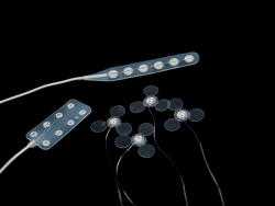
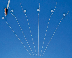
63 What are the characteristics of the three classes of neurons comprising an epileptic focus? Three classes of neurons (Talairach and Bancaud)1: Class I neurons: intrinsically abnormal “epileptic neurons,” characterized by paroxysmal depolarization shift (PDS) Class II neurons: labile neurons, which, depending on their “physiological occupation,” may discharge with a normal pattern or an abnormal burst index. (They can be “recruited” by class I neurons to discharge abnormally. If occupied with appropriate physiological stimulus, they can resist recruitment into abnormal activity.) Class III neurons: neurons with a normal burst index What portion of patients with epilepsy are seizure-free on their first antiepileptic drug (AED)? 47% (seizure-free in this context is defined as having had no seizure in a 1-year period)2 What portion of patients with epilepsy are seizure-free on their second antiepileptic drug (AED)? 13% (seizure-free in this context is defined as having had no seizure in a 1-year period)2 What portion of patients with epilepsy are refractory to medical therapy? 40%2 Yes. Patients in whom ≥20 seizures have occurred are less likely to respond to AEDs. What are the phases in preoperative workup of patients with epilepsy? Phase I: • Imaging • Scalp EEG/video EEG (recorded video synchronized with EEG) • Neurophysiological testing Phase II: • Invasive monitoring • Subdural grids and strips • Depth electrodes • Wada test What imaging methods are useful in the characterization of the seizure focus? 1. Interictal MRI: to assess abnormalities of brain structure. It is sensitive and specific for neoplasia, dysplasia, vascular malformations, and hippocampal sclerosis (gold standard for diagnosis and for quantifying volume). 2. Interictal proton magnetic resonance spectroscopy (MRS): to study reductions in N-acetylaspartate (NAA) as a measure of neuronal integrity (by comparing NAA levels with choline or creatine). A decreased NAA-to-choline ratio is found in lesions such as hippocampal sclerosis (HS). 3. Ictal/interictal single-photon emission computed tomography (SPECT): to characterize cerebral perfusion during and after a seizure, with subtraction of ictal-interictal images. It is sensitive for lateralizing seizure focus. 4. Interictal FDG positron emission tomography (PET): a. With FDG (fluoro-2-deoxyglucose) to study metabolism. Anterior temporal lobe hypometabolism and extratemporal hypometabolism provide accurate lateralization in temporal lobe epilepsy (TLE). b. With flumazenil to study density of central benzodiazepine receptors (cBZRs) 5. Magnetoencephalography (MEG): to detect small electrical currents produced by neurons. MEG is free of distortions caused by the scalp and cranium as well as by irregularities secondary to masses and previous surgery. What are the major limitations of surface EEG recordings (in comparison to invasive monitoring)? 1. Scalp EEG is less spatially precise than invasive monitoring. 2. Scalp EEG signals have a lower amplitude and bandwidth (filtered by skull, CSF, dural layers) and are less sensitive in the early detection of abnormal neural activity characterizing the onset of a seizure. 3. Scalp EEG recordings contain more noise, including muscle and movement artifact, each of which can obscure signals of interest. What is the purpose of the Wada test, and who developed it? The Wada test is used to assess language and memory localization. It was developed by Juhn Wada in 1949 to determine hemispheric dominance for language.3 What is another name for the Wada test? Intracarotid sodium amobarbital test (IAT) What intervention is performed during the Wada? Injection of sodium amobarbital into the ICA What are some semiinvasive EEG recording electrodes? Semiinvasive chronic recording electrodes include: 1. Foramen ovale (FO) electrodes 2. Peg electrodes4 What are the invasive electrodes used in EEG recording? 1. Subdural strip and grid electrodes 2. Depth electrodes How are foramen ovale electrodes placed? Foramen ovale electrodes are placed using the same techniques used for placement of a lesioning electrode in the treatment of trigeminal neuralgia (discussed in Chapter 62). What is the purpose of foramen ovale electrode placement? Foramen ovale electrodes are used for recording from the mesiobasal temporal lobe (TL) and are therefore helpful in lateralizing seizure onsets to the mesial portion of the TL. Peg electrodes are, as their name implies, peg-shaped electrodes that are inserted into holes in the skull. They provide epidural recordings and are helpful when the precise location of a focus is not yet determined and can be placed to further determine where to implant strip, grid, or depth electrodes. Peg electrodes may also be placed contralateral to these other conventional electrodes to rule out contralateral seizure activity.5 What are depth electrodes? Depth electrodes are cylindrical electrode arrays with a diameter of slightly more than 1 mm that are implanted into deep nuclei and structures, usually the amygdala and hippocampus. How are depth electrodes typically placed? Depth electrodes are implanted using stereotactic techniques through a burr hole. The hippocampus, a common recording target, is often approached laterally (orthogonally) via a burr hole in the temporal bone (more common) or posteriorly (longitudinally) via a burr hole in the occipital bone.6 What are subdural strip electrodes? Subdural strip electrodes are linear arrays of electrodes embedded in a flexible silicone strip, with each electrode electrically connected to a wire that emanates as a conductor within a wire bundle from one side. What type of activity are subdural strip electrodes often used to monitor? Subdural strip electrodes are used to monitor neocortical activity. How are subdural strip electrodes placed? Subdural strip electrodes are commonly placed using a burr hole. Stereotactic navigation may be used to guide electrode placement. They are advanced in the subdural space, and by virtue of the flexibility of the silicone, conform to the brain surface. Where are subdural strip electrodes placed for monitoring of the temporal lobe? Three are often used: 1. An anterior temporal electrode is inserted such that the distal electrode curves around the tip of the temporal lobe. 2. A middle temporal electrode is inserted to curve along the inferior surface of the temporal lobe, with the most distal contact overlying the parahippocampal gyrus. 3. A posterior temporal electrode is inserted posteriorly to course along the middle temporal gyrus and to pass posterior to the end of the sylvian fissure. What electrodes are shown in Fig. 63.1? Fig. 63.1 (Courtesy of Ad-Tech. Reprinted by permission.) 1. Subdural strip electrodes (top, 1 × 6 array) 2. Subdural grid electrodes (left, small 2 × 4 array) 3. Intraoperative (IOM) mapping electrode (bottom right, single electrode with cloverleaf tab design for stabilization during mapping) What electrodes are shown in Fig. 63.2? Fig. 63.2 (Courtesy of Ad-Tech. Reprinted by permission.)
Epilepsy Surgery
63.1 Pathophysiology: Seizure Focus
63.2 Preoperative Workup


Epilepsy Surgery
Only gold members can continue reading. Log In or Register to continue

Full access? Get Clinical Tree


