Figure 18-1 Right radial motor nerve conduction studies, demonstrating conduction block when the nerve was stimulated at the axilla and Erb’s point, recording over the extensor digitorum communis. “Sp. Grove” (sic) indicates stimulation site in the distal upper arm over the spiral groove, distal to the site of the patient’s bruise.
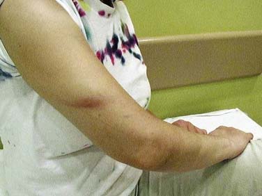
The presence of partial conduction block and the finding of reduced recruitment yet normal motor unit potential morphology in clinically weak muscles were difficult to reconcile with the reported 4-month history of right wrist and finger drop. We believed that the patient’s clinical picture and electrodiagnostic studies were inconsistent with a 4-month-old compressive radial neuropathy, or for multifocal motor neuropathy or other chronic, acquired demyelinating polyneuropathy. This prompted us to perform a complete neuromuscular evaluation, at which time we noticed a bruise of the right upper arm over the spiral groove (Fig. 18-2) that had been overlooked by us during the EMG procedure. When she was questioned about this bruise she told us that she had fallen asleep overnight at her computer desk a “few days” before her visit with us. However, she maintained that there had been no change in wrist and finger function relative to that event. Integrating the history, examination, and electrodiagnostic testing, however, led us to conclude that most, if not all, of the patient’s current weakness was related to the second compressive injury. The patient acknowledged our clinical impression and asked us to support her long-term disability claim.
CONCLUSION
The two most common causes of radial neuropathy are humeral fracture and external compression during deep sleep (i.e., “Saturday night palsy”). Radial neuropathies associated with bone fractures most commonly occur with humeral fractures of the upper arm, although they can occur with fractures of the radius or ulnar in the forearm and wrist.1 There are several potential mechanisms of nerve injury associated with bone fracture: direct damage from the blunt trauma that also fractures bone; laceration, compression, or stretching caused by bone fragments; and injury during efforts to reduce the fracture.2 In acute compressive radial neuropathies, the nerve may be compressed against the medial aspect of the humerus (e.g., caused by the head of a sleeping partner or draping the arm over a chair for prolonged periods) or against the lateral aspect of the humerus (e.g., sleeping on the side with the arm underneath the body pressed against a hard surface); the latter, we believe, being the mechanism and site of injury for our patient.
In acute compressive radial neuropathies, nerve conduction studies typically demonstrate conduction block and slowing at the site of compression,3 indicative of focal demyelination. These abnormalities resolve within 6 to 8 weeks, in parallel to clinical improvement. In contrast, patients with traumatic nerve injury, for example, caused by fracture, are more likely to demonstrate conduction failure on motor and sensory studies, indicative of more severe nerve injury. In these traumatic cases, recovery of motor and sensory function is more protracted and often incomplete.1,3,4,5
When nerve tissues are subjected to external compression, some of the compressed tissue is displaced to sites of lower pressure. The sequential histopathologic events of acute compressive neuropathy are (1) expression of endoneurial fluid, (2) expression of fluid within axons, (3) expression of cytoskeletal elements, and (4) distortion and disruption of myelin and Schwann cell elements.6,7 Nodes of Ranvier frequently become obscured and lengthened because of displaced paranodal myelin, and these changes contribute to the loss of saltatory conduction.6,8 Focal ischemia and endoneurial edema may also contribute to the focal pathology.9
In our case, the upper arm bruise, radial nerve conduction findings of focal conduction block near the contusion site, and the needle EMG findings of reduced recruitment with normal motor unit morphology argued that the patient’s radial neuropathy was caused by acute compression rather than a chronic process or an old, acute-onset injury without recovery. This change of estimated duration of radial neuropathy improved prognosis (favorable, if further compression is avoided) and suggested that surgical exploration at that time was probably not indicated.
Related posts:
 Malignant Peripheral Nerve Sheath Tumor
Disseminated Sporotrichosis with Multiple Granulomatous Mononeuropathies
Adrenomyeloneuropathy Masquerading as Charcot-Marie-Tooth Disease
Focal Mononeuropathy Onset of Amyotrophic Lateral Sclerosis
A Case of Guillain-Barré Syndrome Associated with Anti-GD1b Immunoglobulin G Antibodies
Length-Related Axonal Loss in Neuropathy
Malignant Peripheral Nerve Sheath Tumor
Disseminated Sporotrichosis with Multiple Granulomatous Mononeuropathies
Adrenomyeloneuropathy Masquerading as Charcot-Marie-Tooth Disease
Focal Mononeuropathy Onset of Amyotrophic Lateral Sclerosis
A Case of Guillain-Barré Syndrome Associated with Anti-GD1b Immunoglobulin G Antibodies
Length-Related Axonal Loss in Neuropathy
Stay updated, free articles. Join our Telegram channel

Full access? Get Clinical Tree


