Figure 80-1 Illustration of the process of amyloid specific microdissection. A, Peripheral nerve biopsy stained with Congo red indicating amyloid positive regions (arrows). B, Amyloid-positive areas are selected and outlined for laser microdissection (arrows). C, Laser specifically removes selected area (arrows). Inset illustrates the tissue collected into microfuge cap for processing (arrow).
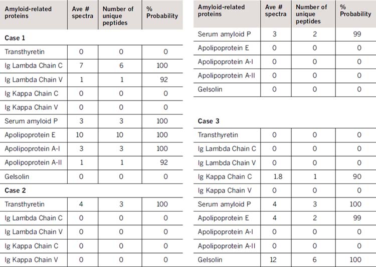
Table 80-1 illustrates the utility of this method using three different patient cases from peripheral nerve biopsy specimens. A list of amyloid-related proteins is indicated in Column 1. Values indicate the average number of MS spectra, the number of unique peptides observed, and the protein identification probability for each type of amyloid protein observed. Values of two or more spectra in more than one dissection sample, with greater than 90% protein identification probability indicates positive protein identification for the specimen. Test results from Case 1 indicate AL type amyloid, results from Case 2 indicate TTR type amyloid, and the results from Case 3 suggest the rare Gelsolin type amyloid. Each case was previously characterized for amyloid type by clinical history and immunohistochemistry using a panel of antibodies against amyloid proteins (TTR, SAA, lambda and kappa light chains and SAP).
Related posts:
 Malignant Peripheral Nerve Sheath Tumor
Disseminated Sporotrichosis with Multiple Granulomatous Mononeuropathies
Late Sporadic CMT4C—A New KIAA1985 Mutation
Late-Onset Transthyretin Val30Met Familial Amyloid Polyneuropathy Unrelated to Endemic Foci
A Weak, and Numb Patient with Tremor—Antimyelin-Associated Glycoprotein Polyneuropathy
Copper Deficiency Myeloneuropathy
Malignant Peripheral Nerve Sheath Tumor
Disseminated Sporotrichosis with Multiple Granulomatous Mononeuropathies
Late Sporadic CMT4C—A New KIAA1985 Mutation
Late-Onset Transthyretin Val30Met Familial Amyloid Polyneuropathy Unrelated to Endemic Foci
A Weak, and Numb Patient with Tremor—Antimyelin-Associated Glycoprotein Polyneuropathy
Copper Deficiency Myeloneuropathy
Stay updated, free articles. Join our Telegram channel

Full access? Get Clinical Tree


