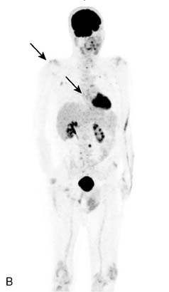
Figure 57-1 A, F-18 fluorodeoxyglucose positron emission tomography scan reveals hypermetabolic lesions in the right humerus and thoracic (T9) vertebral body, on maximal intensity projection images. B, Improvement in the hypermetabolic activity after radiation therapy.
He was treated with radiation to the right humeral head and T9. His burning resolved and tingling sensations in the feet resolved. He had improvement, with increased ankle strength (previously MRC 0). He had decreased sensation to pinprick, in the toes and feet, which was previously absent. He is able to walk without a cane. His edema was improved. His VEGF is reduced to 336 pg/mL (31-86). A repeat PET scan noted reduced hypermetabolism (see Fig. 57-1B) and a reduction in the splenomegaly.
CONCLUSION
Osteosclerotic myeloma is a plasma cell dyscrasia, characterized by sclerotic bone lesions and a progressive demyelinating neuropathy.1,2 Osteosclerotic myeloma may be associated with other systemic manifestations, and the term POEMS syndrome designates patients with a polyneuropathy, organomegaly, endocrinopathy, m-protein, and skin changes. Many patients do not have all of these features, and the osteosclerotic myeloma and edema, which are not in the eponym, are frequent.1
The osteosclerotic myeloma is typically detected on a skeletal survey, which is preferable to a bone scan. The osteosclerotic lesion may be subtle and difficult to detect. Patients with a monoclonal gammopathy and a demyelinating neuropathy should have a skeletal survey, which is more sensitive than a bone scan, to detect osteosclerotic lesions.2 Our patient did not have an osteosclerotic lesion detected on skeletal survey.
The patient also had received etanercept and acquired demyelinating neuropathies have occurred following the use of this and other tumor necrosis factor antagonists.3 In previous reports of this neuropathy, some patients improve when etanercept is stopped; however, others were treated successfully with typical medications for CIDP. The patient did have a fat pad aspirate that was positive for amyloid on Congo red staining. This was thought to be a false-negative finding because the open abdominal fat pad biopsy, nerve biopsy, and bone marrow biopsy were all negative for amyloid. Congo red staining for amyloid on fat pad aspirates may be nonspecific.4
Because the typical syndrome includes a demyelinating neuropathy, a faint lambda monoclonal protein, and edema, the diagnosis of POEMS was pursued. The patient did have an elevated VEGF, so despite a negative skeletal survey, a FDG-PET scan identified a hypermetabolic lesion, correlating with an osteosclerotic lesion seen on the computed tomography scan, not previously identified (see Fig. 57-1A). The FDG-PET scan was also helpful in following a response to treatment with radiation (see Fig. 57-1B).
VEGF may play a role in the pathogenesis, and can be measured in the serum and used as a diagnostic marker.5–7 The diagnosis of POEMS or osteosclerotic myeloma can be difficult. Some patients with POEMS are initially diagnosed with CIDP and are only diagnosed later after a poor response to conventional immunomodulatory treatment for CIDP. Radiation, surgical removal of the plasmacytoma, or autologous peripheral blood stem cell transplant may result in improvement of the neuropathy.8,9
Related posts:
 Malignant Peripheral Nerve Sheath Tumor
Disseminated Sporotrichosis with Multiple Granulomatous Mononeuropathies
Late Sporadic CMT4C—A New KIAA1985 Mutation
Late-Onset Transthyretin Val30Met Familial Amyloid Polyneuropathy Unrelated to Endemic Foci
A Weak, and Numb Patient with Tremor—Antimyelin-Associated Glycoprotein Polyneuropathy
Copper Deficiency Myeloneuropathy
Malignant Peripheral Nerve Sheath Tumor
Disseminated Sporotrichosis with Multiple Granulomatous Mononeuropathies
Late Sporadic CMT4C—A New KIAA1985 Mutation
Late-Onset Transthyretin Val30Met Familial Amyloid Polyneuropathy Unrelated to Endemic Foci
A Weak, and Numb Patient with Tremor—Antimyelin-Associated Glycoprotein Polyneuropathy
Copper Deficiency Myeloneuropathy
Stay updated, free articles. Join our Telegram channel

Full access? Get Clinical Tree


