• The patient is placed into a direct lateral position.
• It is essential that the patient be directly perpendicular to the ground to ensure true anteroposterior (AP) and lateral fluoroscopic views of the disk space.
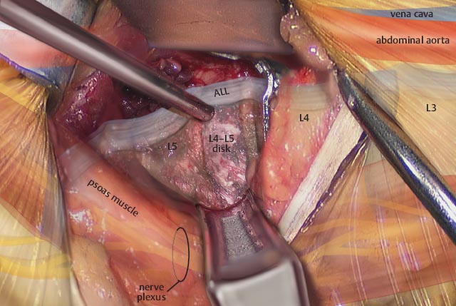
• AP and lateral fluoroscopic imaging is used to identify the disk space.
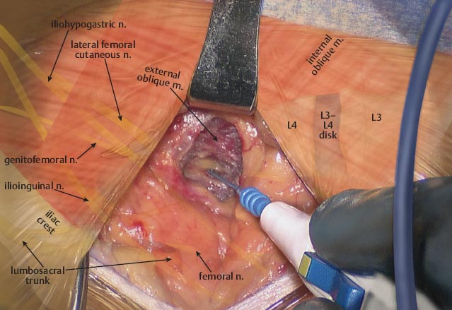
• A skin incision is made, exposing the fibers of the external oblique muscle.
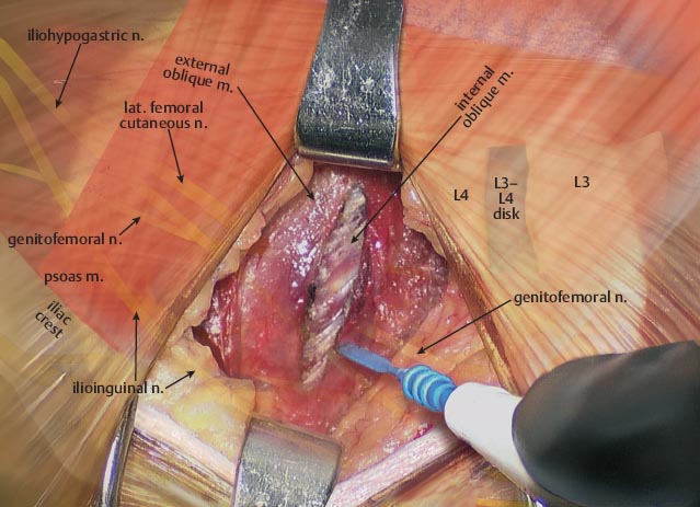
• The fibers of the internal oblique muscle lie underneath the external oblique.
– These fibers run in an opposite direction.
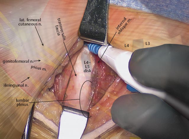
• Once the fibers of the internal oblique are split, the transversalis fascia is identified.
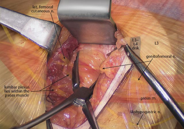
• The transversalis fascia is bluntly opened, exposing the retroperitoneal fat.
Related posts:


Stay updated, free articles. Join our Telegram channel

Full access? Get Clinical Tree


