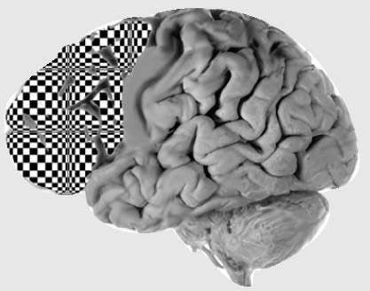
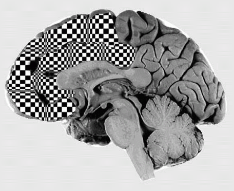
As the most voluminous portion of the brain’s four lobes, the frontal lobes continue to myelinate throughout the second decade. The disproportionate size of the adult human frontal lobe compared with that of other mammals or even children reinforces the idea that the frontal lobes facilitate living in a socially demanding environment. Impairment of functions such as planning, complex decision making, and inhibiting inappropriate impulses are the functions most relevant to this review.
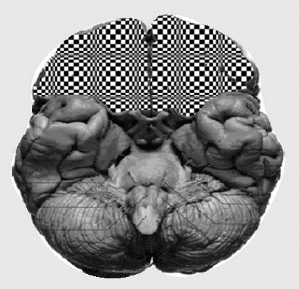
A useful approach differentiates the function of three main anatomical regions of the cortex using an anterior view of the brain. In practice, many patients may have symptoms referable to more than one of these three regions or may display only one of the symptoms, suggestive of a single region (Table 7.1).
Diseases affecting the white matter of the frontal lobes may also demonstrate combinations of the clinical characteristics listed previously. In addition to frontotemporal dementia, a degenerative disorder directly impairing frontal gray matter, the differential of such illnesses is broad and includes (but is not limited to) multiple sclerosis, chronic hydrocephalus, and vascular disease. Degenerative diseases affecting the frontal lobes are discussed further in Chapter 9, The Dementias.
Supportive signs and symptoms suggesting frontal lobes dysfunction should be sought and documented whether present or absent. These include Broca’s aphasia on the left and occasionally aprosodia on the right, frontal eye field involvement (hemineglect, gaze deviation/preference away from lesion), and, in larger lesions, hemiparesis.
Pathological reflexes seen in frontal lesions (frontal release signs) are easily elicited with practice. These include the grasp (a tendency to squeeze anything placed in the palm), palmomental (scratching the palm from wrist toward thumb causes a contraction of the mentalis on the same side), snout (tapping the lips leads to a pursing of the mouth), and glabellar reflexes (an inability to suppress closing the eyes when forehead is tapped).
▪ PARIETAL LOBE FUNCTION
Parietal lobe damage can produce an interesting array of clinical deficits and, because several parietal functions are highly lateralized, they are exceptionally helpful for localization. In this section we’ll discuss focal syndromes, though it should be noted that more diffuse processes (e.g., Alzheimer’s disease) can preferentially affect the parietal lobes.
Aphasia is an acquired disorder of language and is often highly localizable (Fig. 7.1). Language impairments should affect all output modalities (writing as well as speech, for example). In the general population about 95% to 99% of right-handers and 70% of left-handers have left cerebral dominance of language, placing language function in the left hemisphere in about 95% of people (depending on which study you reference).
Major features: Poor response inhibition (perseveration) Environmentally bound behaviors Impairments in: Working memory and executive function Sustained attention Set shifting Judgment
OFC (emotional modulation)
Trauma (associated anosmia) Tumors FTD
Observation
Rating scales
Cognitive testing often normal
Major features: “Pseudopsychopath” Syndrome of acquired sociopathy: tactless, inappropriate, impulsive Easily distracted, lability of emotional regulation Impairments in: Knowing social boundaries Controlling high-risk behavior
mPFC (initiation and monitoring)
NPH ACA strokes Hydrocephalus Tumors
Acute-unilateral Chronic-bilateral
Stroop observation
Major features: Bradyphrenia Abulia: apathy and impaired initiation Decreased motor, cognitive, and emotional activity Akinetic mutism (coma vigil): most severe form of abulia Complete absence of initiation—patients appear awake and alert but have a paucity of movement and essentially no verbal output
ACA, anterior cerebral artery; DLPFC, dorsolateral prefrontal cortex; mPFC, medial prefrontal cortex (includes anterior cingulate and supplementary motor area); OFC, orbital frontal cortex.
Only gold members can continue reading. Log In or Register to continue