Sleep Disorders


Sleep Disorders


Jean K. Matheson


▪ INTRODUCTION

Sleep complaints accompany most psychiatric disorders. Conversely, primary sleep disorders may mimic, exacerbate, or possibly induce psychiatric disease. Sleep disorders may represent a primary disorder of mechanisms regulating sleep or failure of a specific organ system manifesting in a unique way during sleep. Sleep complaints should not be ignored or treated empirically with pharmacological agents without analysis of the etiology.

In practice, some aspects of sleep physiology can be objectively studied with polysomnography (PSG), the simultaneous recording of electroencephalographic, cardiopulmonary, and motor parameters during sleep. The classification of sleep disorders is based on both clinical and physiological criteria; the current classification of sleep disorders is outlined in the International Classification of Sleep Disorders Diagnostic and Coding Manual second edition (ISCD-2), a product of a task force of the American Academy of Sleep Medicine. The major diagnostic categories are outlined in Table 21.1.


▪ OVERVIEW OF SLEEP


Organization of Sleep Stages

Rapid eye movement (REM) sleep, sometimes called dreaming sleep, and non-REM (NREM) sleep are the two sleep states. REM sleep alternates with NREM in recurring cycles of approximately 90 minutes. Recordings derived from electroencephalography (EEG), eye movements (electro-oculograms [EOGs]), and surface electromyography (EMG) of muscles (typically chin) are necessary to identify sleep states. NREM sleep has classically been divided into four stages (1 to 4), which represent progressive deepening of sleep. A recent revision of staging nomenclature now classifies these stages as N1, N2, and N3 (
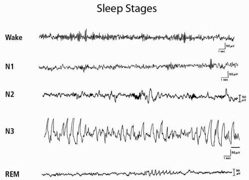
Fig. 21.1). The waking EEG in quiet wakefulness shows the characteristic alpha rhythm, a posterior predominant 8- to 13-Hz rhythm that attenuates with eye opening. Stage N1 (stage 1) is characterized by the gradual disappearance of alpha rhythms, which are replaced by 4- to 7-Hz theta rhythms and some faster activity. The emergence of sleep spindles (11- to 16-Hz sinusoidal transients lasting at least 0.5 seconds) and K complexes (negative sharp wave followed by positive component lasting ≥0.5 seconds) defines N2 (stage 2). Stages 3 and 4 are now grouped together under the term N3. N3 is characterized by high-voltage slow wave activity with frequencies of 0.5 to 2 Hz. Stage N3 is often referred to as delta sleep, slow wave sleep, or deep sleep. N3 can be characterized as “deep sleep” because subjects are difficult to arouse and typically amnestic for events that occur on arousal from this sleep stage. Although detailed narrative dreaming does not seem to occur, subjects awoken from N3 may report fragmentary dream mentation or that they were “thinking” (see parasomnias below). The normal young adult descends in an orderly progression through the three NREM stages. N3 appears approximately 30 to 40 minutes after sleep onset. The first REM period (stage R) follows this slow wave sleep, approximately 70 to 90 minutes after sleep onset.


TABLE 21.1 INTERNATIONAL CLASSIFICATION OF SLEEP DISORDERS-2 (ICSD-2) DIAGNOSTIC CATEGORIES


I.


Insomnia


II.


Sleep-Related Breathing Disorders


III.


Hypersomnias of Central Origin Not Due to a Circadian Rhythm Sleep Disorder, Sleep Related Breathing Disorder, or Other Cause of Disturbed Nocturnal Sleep


IV.


Circadian Rhythm Sleep Disorders


V.


Parasomniass


VI.


Sleep-Related Movement Disorders


VII.


Isolated Symptoms, Apparently Normal Variants, and Unresolved Issues


VIII.


Other Sleep Disorders


The PSG during REM sleep shows dramatic changes. A sudden loss of EMG activity occurs in the chin muscles, which is indicative of generalized skeletal muscle atonia. Rapid eye movements occur in phasic bursts, and the EEG shows mixed frequencies similar to those in waking and stage 1 sleep, sometimes with a characteristic “saw tooth” pattern.

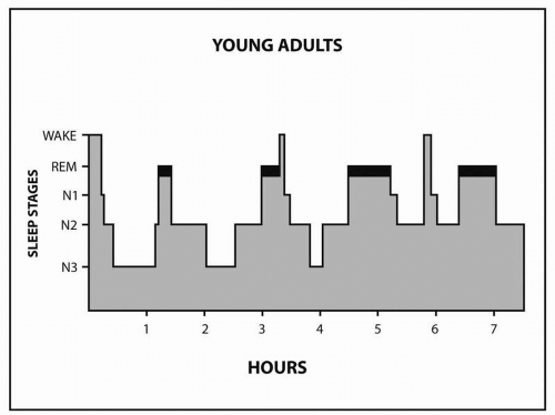
The first REM period is short, lasting about 10 minutes. The end of the first REM period completes the first sleep cycle. Thereafter, NREM sleep continues to alternate with REM sleep; the healthy adult goes through 4 to 6 cycles (Fig. 21.2).

Sleep architecture is the organization of sleep stages and cycles. The normal young adult spends approximately 5% of the night in stage N1, 50% to 55% in stage N2, 20% in N3, and 20% to 25% in REM. N3 is concentrated in the first third of the night, whereas REM episodes become progressively longer later in the night. N3 decreases as a function of age, whereas REM remains fairly constant after early childhood. Of the newborn’s daily 17 to 18 hours of sleep, 50% is REM. Children and early adolescents sleep 10 to 11 hours. Most adults prefer to sleep 7.5 to 8.5 hours. Subjects sleep-restricted to 6 hours show cognitive deficits that are cumulative. Deprivation of REM sleep by medication, sleep disruption, or sleep deprivation results in REM rebound when the cause of the deprivation is removed. Sleep deprivation also induces N3 rebound sleep during recovery sleep.


FIGURE 21.1 Sleep stages.


FIGURE 21.2 Hypnogram in a normal young adult.

REM and NREM sleep differ physiologically. REM sleep is characterized by both phasic and tonic changes in physiology. The drop in baseline EMG correlates with a tonic change. Rapid eye movements correlate with phasic changes. Tonic physiological changes also include impaired thermoregulation, reduction in ventilatory chemosensitivity, hypotension, bradycardia, increased cerebral blood flow and intracranial pressure, increased respiratory rate, and penile erection. Phasic changes include vasoconstriction, increased blood pressure, tachycardia, and further increases in cerebral blood flow and respiratory rate. During NREM sleep, the physiological state is more stable, with an overall reduction in blood pressure, heart rate, cardiac output, and ventilation. One characteristic feature of N3 is the secretion of growth hormone.

Some disorders are exacerbated by or occur only during certain sleep stages. Sleepwalking, for example, occurs with arousal from NREM, usually N3 sleep. Epileptic seizures tend to be facilitated by NREM sleep but inhibited by REM sleep. Obstructive sleep apnea is typically worse in REM sleep because of REM atonia and decreased respiratory chemosensitivity.


▪ NEUROBIOLOGY OF SLEEP

There are two sleep drives, one homeostatic (called process S) and the other circadian (process C). A homeostatic drive increases with time spent awake, and the circadian drive is under the influence of the suprachiasmatic nucleus (SCN) of the hypothalamus (see later discussion).
Sleep and wake states represent a complex interaction between wake-promoting arousal and sleep-promoting networks. The ascending arousal system comprises discrete cell groups and their projecting axons originating in brainstem, hypothalamus, and basal forebrain. Neurons of the ventrolateral preoptic nucleus (VLPO) of the hypothalamus are sleep active and sleep promoting and innervate wake-promoting areas, including the neurons of the posterolateral hypothalamus, histaminergic neurons of the tubomammillary nucleus, dopaminergic neurons of the ventral tegmental area, the serotonergic dorsal raphe, norepinephrine-containing neurons of the locus ceruleus, and the cholinergic neurons of the dorsal midbrain and pons. In turn, monoaminergic and cholinergic wake-promoting areas inhibit VLPO, thereby resulting in reciprocal inhibitory relationships that self-reinforce stable periods of sleep and wake. In this model, homeostatic and circadian drives are hypothesized to shift the balance between states by still unknown mechanisms. Adenosine, which accumulates during wakefulness, and whose effect is antagonized by caffeine, may be one of the factors that signals the homeostatic drive to sleep by inhibiting cholinergic arousal systems and activating VLPO. Indirect circadian input into VLPO has been documented.

Brainstem generation of REM sleep is under the influence of the hypothalamus. REM sleep is inhibited by hypocretin-containing cells of the posterior hypothalamus; these cells are lost in narcolepsy.


▪ CIRCADIAN RHYMICITY

Circadian rhythmicity of multiple physiological and behavioral variables, with a period close to 24 hours, is seen in almost all living organisms, even in the absence of environmental cues. Sleep, cortisol secretion, core body temperature, and melatonin secretion by the pineal gland are examples of these rhythms. This internal rhythmicity has long suggested an endogenous clock that can be synchronized by external cues, especially light. The hypothalamic SCN, which receives direct and indirect input from the retinohypothalmic tract, is the major anatomical location of this circadian pacemaker. Intrinsically photosensitive retinal ganglion cells contain the light-sensitive pigment melanopsin, depolarize maximally in response to blue light, and provide the major input into SCN. Rods and cones contribute, but are not required for photic influence of SCN. Melatonin and core body temperature are both good markers of circadian timing (phase) of the SCN. Melatonin levels measured in constant dim light are low during the day; begin to increase in the evening, approximating dusk; are maximal during the night; and abruptly decline in the morning near dawn. Melatonin is secreted by the pineal under the direct influence of the SCN through a circuitous pathway from the hypothalamus to the intermediolateral cell column of the spinal cord, to the superior sympathetic ganglion and finally the pineal. Light serves to activate SCN and immediately turn off melatonin production.
A circadian temperature nadir approximately 2 to 3 hours before a subject’s usual awakening is assigned by convention—circadian time zero, which also correlates with peak melatonin levels. Appropriately timed light exposure can shift the phase of the endogenous melatonin and core body temperature rhythm within 2 to 3 days. By way of a feedback loop involving melatonin receptors on the SCN, appropriately timed melatonin can also shift circadian rhythms. Light in the evening delays rhythms, and light in the morning induces phase advances; conversely, melatonin in the afternoon or evening advances rhythms and morning melatonin results in delay. These effects have important clinical implication, as described in the section on circadian disorders. The phase response curve for light, which indicates the degree and direction of phase change at any given time in the circadian day, is well established, but the phase response curve for melatonin is still not completely resolved. The period of the human circadian pacemaker is approximately 24.2 hours.


▪ SLEEP CLINICAL NEUROPHYSIOLOGY


Polysomnography

PSG is the term applied to the simultaneous and continuous measurement of multiple physiological parameters during sleep. In practice, the term PSG has come to mean a specific type of polysomnographic study in which measurements allow for the identification of sleep stage, monitoring of cardiopulmonary function, and monitoring of body movements during sleep. This study is typically obtained at night in a sleep laboratory for the purpose of identifying, as best as possible given the novel environment, the patient’s typical sleep and its associated pathologies. The multiple sleep latency test (MSLT) and the maintenance of wakefulness test (MWT) are more limited daytime sleep studies that are useful in the evaluation of narcolepsy and other causes of daytime sleepiness. The MSLT measures the tendency to fall asleep during the day and screens for the occurrence of inappropriate daytime episodes of REM sleep during multiple daytime naps. The MWT is the inverse of the MSLT and measures the ability to stay awake in multiple daytime naps. Parameters recorded in standard PSG, MSLT, and MWT are indicated in Table 21.2.

The American Academy of Sleep Medicine (AASM) has developed guidelines for the indication for polysomnography. These indications include the following:


  • Suspicion of sleep-related breathing disorders


  • Treatment and follow-up of sleep-related breathing disorders


  • In combination with the MSLT for suspected narcolepsy or idiopathic hypersomnia


  • Evaluation of sleep-related behaviors that are violent, potentially injurious, or do not respond to conventional therapy


  • To assist in the diagnosis of paroxysmal arousals that are suggestive of seizure disorder (with additional video and EEG


  • Evaluation of sleep-related movement disorders.


TABLE 21.2 POLYSOMNOGRAPHY


TEST


PARAMETERS RECORDED


PSG


EEG (F4-M1, C4-M1, O2-M1)


Additional EEG if indicated


Electro-oculogram (EOG) (eye movement)


Electromyography (EMG) (chin)


Airflow


Respiratory effort


Oxygen saturation


Electrocardiogram (ECG)


EMG limb, anterior tibialis muscles; extensor digitorum muscles when indicated


Body position


Esophageal pH (rarely)


MSLT and MWT


EEG (F4-M1, C4-M1, O2-M1)


EOG (eye movement)


EMG (chin)


ECG


Optional: Respiratory monitoring


F4, C4, O2 refer to frontal, central, and occipital EEG leads, respectively, according to the International 10-20 System; M1 refers to a mastoid reference.


Because sleep-related breathing disorders can have broad clinical presentations and implications, PSG may be indicated in many clinical situations, including congestive heart failure, arrhythmia, coronary artery disease, stroke, hypertension, pulmonary disease, neuromuscular disease, headache, and gastroesophageal reflux. Although PSG is not routinely indicated for the evaluation of chronic insomnias, a history suggestive of a contributing sleep disorder, especially sleep-related breathing or movement disorder, does support the use of PSG. On the other hand, despite research interest in the changes in sleep associated with depression, PSG is not indicated for the primary purpose of establishing a diagnosis of depression because there are no abnormalities of sleep architecture specific to that diagnosis.


Sleep Architecture

A timeline of sleep stages during the night is reported as a hypnogram (Fig. 21.2). Quantitative analysis also includes a number of variables (Table 21.3). Sleep architecture may be distorted by specific disorders and is commonly disturbed by medications (Table 21.4).


Respiratory Measures

The recording of airflow, respiratory effort, and oxygen saturation allows for the determination of abnormal breathing patterns during sleep. These abnormalities underlie the sleep-related breathing disorders. A variety of abnormal breathing events exist, including the following:

Apnea: The absence of airflow for at least 10 seconds. There are three types:


  • Obstructive apnea: At least 90% reduction in airflow for at least 10 seconds with evidence of persistent respiratory effort (Fig. 21.3)


  • Central apnea: At least 90% reduction in airflow for 10 seconds without evidence of any respiratory effort (Fig. 21.4)


  • Mixed apnea: At least 90% reduction in airflow for 10 seconds with initial absence of effort followed by a return of respiratory effort before resumption of airflow.


TABLE 21.3 SLEEP ARCHITECTURE PARAMETERS


VARIABLE


ABBREVIATION


DEFINITION


COMMENTS


Time in bed


TIB


The time from lights out until the subject chooses to end the study.


Excessive time in bed may exacerbate insomnias; common in old age.


Total sleep time


TST


Total time the subject actually slept.


Varies with age: Adult: 7-9 hr Children (5-12): 9-11 hr; (0-2 mo): 10.5-18 hr.


Sleep efficiency index


SE


TST/TIB. Often reported as a percentage. This is an important measure of sleep quality.


Age dependent; average normal adult 85% to 95%; <85% suggests sleep disruption.
High efficiency may indicate prior sleep deprivation.
Decreases with excessive time in bed, increased wake during sleep or prolonged latency.


Percentage of each stage


This may be reported as a percentage of the TIB or the TST.


Varies with; age approximate; young adult: 5% stage N1, 50% to 55% stage N2, 20%, stage N3, 20% to 25% stage REM.


Sleep onset latency


SL


Time from lights out until the onset of sleep. Sleep onset is usually defined as the onset to the first epoch of any sleep stage. Sometimes reported as latency to 3 epochs of stage one or one epoch of any other sleep stage (unequivocal sleep).


Less than 20 minutes; prolonged in insomnias and delayed sleep phase.


Number of awakenings


Records the number of times the subject returns to stage wake after sleep onset.


Occasional awakenings are common; increased awakenings occur in association with disorders causing arousal.


Number of arousals


Records the number of EEG arousals. Arousal defined as 3 seconds with increased EEG frequency above baseline without awakening.


Increased in disorders such as sleep apnea and periodic limb movements.


REM latency


Reports time from sleep onset to first REM period.


Average 90 minutes in adults. Lower latencies are reported in depression but are nonspecific. Many drugs prolong REM latencies. REM rebound shortens latency.


EEG, electroencephalogram; REM, rapid eye movement.


TABLE 21.4 MEDICATION EFFECTS ON THE POLYSOMNOGRAM


DRUGS


SWS


REM


MISCELLANEOUS


TCAs


imageimage


SSRIs and SNRIs


⇔?


image


SSRIs and SNRIs image non-REM slow eye movements


Nefazodone


image


Trazodone


image


imageimage


Bupropion


image


NE and DA uptake inhibitor


Mirtazapine


MAOIs


imageimageimage


Lithium


image


image


BZDs


image


image


image Spindle activity and stage 2


Zolpidem


Dopaminergic drugs


?


?


Mixed results


Anticonvulsants


Minimal data


Phenytoin


image


?


Carbamazepine


image


image


Tiagabine


image


Gabapentin


image


image


Lipophilic beta-blockers


image


image


Clonidine


?


image


Opioids


image


image


Can image respiratory drive


Amphetamines


image


image


Caffeine


image


?


Adenosine antagonist
Adenosine image SWS


Alcohol (acute)


image


image


After the metabolism of alcohol there is a REM rebound, often in second half of night Chronic use image SWS and REM


Sodium oxybate (GHB)


image


imageimage


Approved for cataplexy and EDS


BZDs, benzodiazepines; DA, dopamine agonist; EDS, excessive daytime sleepiness; GHB, gamma hydroxybutyrate; NE, norepinephrine; REM, rapid eye movement; SNRIs, selective norepinephrine reuptake inhibitors; SSRIs, selective serotonin reuptake inhibitors; SWS, short wave sleep; TCA, tricyclic antidepressants.


Hypopnea: Abnormal respiratory event lasting at least 10 seconds with at least a 30% reduction in thoracoabdominal movement or airflow compared to baseline and with at least a 4% oxygen desaturation (Fig. 21.5)

Only gold members can continue reading. Log In or Register to continue

Stay updated, free articles. Join our Telegram channel

Sep 7, 2016 | Posted by in PSYCHIATRY | Comments Off on Sleep Disorders

Full access? Get Clinical Tree

Get Clinical Tree app for offline access