1 Fundamental concepts in functional neurology
Introduction
Much of the understanding that we have today of how human neurons function was based on the ‘integrate and fire’ concept formed by Eccles in the 1950s which was developed based on studies of spinal motor neurons (Brock et al. 1952). In this model, spinal motor neurons integrate synaptic activity, and when a threshold is reached, they fire an action potential. The firing of this action potential is followed by a period of hyperpolarisation or refraction to further stimulus in the neuron. This early integrate and fire model was then extrapolated to other areas of the nervous system including the cortex and central nervous system which strongly influenced the development of theories relating to neuron and nervous system function (Eccles 1951).
Early in the 1970s, studies that revealed the existence of neurons that operated under much more complex intrinsic firing properties started to emerge. The functional output of these neurons and neuron systems could not be explained by the existing model of integrate and fire for neuron function (Connor & Stevens 1971).
With this fundamental change in the understanding of neuron function came new understanding of the functional interconnectivity of neuron systems, new methods of investigation, and new functional approaches to treatment of nervous system dysfunction.
With the emergence of any clinical science it is essential that the fundamental concepts and definitions are clearly understood. Throughout the textbook the following concepts and terms will be referred to and discussed frequently so it is essential that a good understanding of these concepts be established in the reader’s mind before moving on to the rest of the text.
This chapter will constitute an introduction to the concepts below, which will be covered in more elaborate detail later in the text.
Central integrative state (CIS) of a neuron
The central integrative state (CIS) of a neuron is the total integrated input received by the neuron at any given moment and the probability that the neuron will produce an action potential based on the state of polarisation and the firing requirements of the neuron to produce an action potential at one or more of its axons.
The physical state of polarisation existing in the cell at any given moment is determined by the temporal and spatial summation of all the excitatory and inhibitory stimuli it has processed at that moment. The complexity of this process can be put into perspective when you consider that a pyramidal neuron in the adult visual cortex may have up to 12 000 synaptic connections, and certain neurons in the prefrontal cortex can have up to 80 000 different synapses firing at any given moment (Cragg 1975; Huttenlocher 1994).
‘And’ pattern neurons only fire an action potential if two or more specific conditions are met. ‘Or’ pattern neurons only fire an action potential when one or the other specific condition is present (Brooks 1984).
The thalamic relay cells exhibit complex firing patterns. They relay information to the cortex in the usual integrate and fire pattern unless they have recently undergone a period of inhibition. Following a period of inhibition stimulus, in certain circumstances, they can produce bursts of low-threshold spike action potentials referred to as post-inhibitory rebound bursts. This activity seems to be generated endogenously and may be responsible for production of a portion of the activation of the thalamocortical loop pathways thought to be detected in encephalographic recordings of cortical activity captured by electroencephalograms (EEG) (Destexhe & Sejnowski 2003).
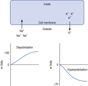
The neuron may be in a state of relative depolarisation, which implies the membrane potential of the cell has shifted towards the firing threshold of the neuron. This generally implies that the neuron has become more positive on the inside and the potential difference across the membrane has become smaller. Alternatively, the neuron may be in a state of relative hyperpolarisation, which implies the membrane potential of the cell has moved away from the firing threshold. This implies that the inside of the cell has become more negative in relation to the outside environment and the potential difference across the membrane has become greater (Ganong 1983) (Fig. 1.1).

Figure 1.1 The effects of ionic movement across the neuron cell membrane. The left side of the diagram illustrates the depolarising effect of sodium ion movement into the cell. The right side of the diagram illustrates the hyperpolarising effect of potassium movement out of the cell. The graphs illustrate the change in potential voltage inside the cell relative to outside the cell as the respective ions move across the membrane. Note the equilibrium potentials for sodium and potassium, +60 and −70 mV, respectively, are reached when the chemical and electrical forces for each ion become equal in magnitude.
The membrane potential is established and maintained across the membrane of the neuron by the flux of ions; usually sodium (Na), potassium (K), and chloride (Cl) ions are the most involved although other ions such as calcium can be involved with modulation of permeability. The movement of these ions across the neuron membrane is determined by changes in the permeability or ease at which each ion can move through selective channels in the membrane.
When Na ions move across the neuron membrane into the neuron, the potential across the membrane decreases or depolarises due to the positive nature of the Na ions, which increases the relative positive charge inside the neuron compared to outside the neuron. When Cl ions move into the neuron, the neuron the membrane potential becomes greater or hyperpolarises due to the negative nature of the Cl ions, which increase the relative negative charge inside the neuron compared to outside the neuron. The same is true when K ions move out of the neuron due to the relative loss of positive charge that the K ions possess.
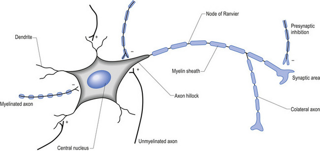
The firing threshold of the neuron is the membrane potential that triggers the activation of specialised voltage gated channels, usually concentrated in the area of the neuron known as the axon hillock or activation zone, that allow the rapid influx of Na into the axon hillock area, resulting in the generation of an action potential in the axon (Stevens 1979) (Fig. 1.2).

Figure 1.2 Anatomical characteristics of a healthy neuron. The central nucleus is maintained by microtubule and microfilament production which requires active protein synthesis. The myelin sheath is composed of oligodendrogliocytes in the central nervous system and Schwann cells in the peripheral nervous system. Note the different types of synaptic contacts illustrated from left to right: axodendritic, axosomatic, dendrodendritic, axohillonic, axoaxonic (presynaptic).
Central integrative state of a functional unit of neurons
The concept of the CIS described above in relation to a single neuron can be loosely extrapolated to a functional group of neurons. Thus, the central integrative state of a functional unit or group of neurons can be defined as the total integrated input received by the group of neurons at any given moment and the probability that the group of neurons will produce action potential output based on the state of polarisation and the firing requirements of the group.
The concept of the central integrative state can be used to estimate the status of a variety of variables concerning the neuron or neuron system such as:
Transneural degeneration
The central integrative state of a neuron or neuron system is modulated by three basic fundamental activities present and necessary in all neurons.
1. Adequate gaseous exchange, namely oxygen and carbon dioxide exchange—this includes blood flow and anoxic and ischaemic conditions that may arise from inadequate blood supply;
2. Adequate nutritional supply including glucose, and a variety of necessary cofactors and essential compounds; and
3. Adequate and appropriate stimulation in the form of neurological communication, including both inhibition and activation of neurons via synaptic activation—synaptic activation of a neuron results in the stimulation and production of immediate early genes and second messengers within the neuron that stimulate DNA transcription of appropriate genes and the eventual production of necessary cellular components such as proteins and neurotransmitters.
Although other activities of neuron function require certain components of oxygen or nutritional supplies, the major necessity of adequate gaseous exchange and adequate nutritional intake into the neuron is to supply the mitochondrial production of adenosine triphosphate (ATP).
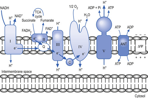
The mitochondria utilise a process called chemiosmotic coupling to harness energy from the food obtained from the environment for use in metabolic and cellular processes. The energy obtained from the tightly controlled slow chemical oxidation of food is used by membrane-bound proton pumps in the mitochondrial membrane that transfer H ions from one side to the other, creating an electrochemical proton gradient across the membrane. A variety of enzymes utilise this proton gradient to power their activities including the enzyme ATPase that utilises the potential electrochemical energy created by the proton gradient to drive the production of ATP via the phosphorylation of adenosine diphosphate (ADP) (Alberts et al. 1994). Other proteins produced in the mitochondria utilise the proton gradient to couple transport metabolites in, out of, and around the mitochondria (Fig. 1.3).

Figure 1.3 Enzymes of oxidative phosphorylation. Electrons (e−) enter the mitochondrial electron transport chain from donors such as reduced nicotinamide adenine dinucleotide (NADH) and reduced flavin adenine dinucleotide (FADH2). The electron donors leave as their oxidised forms, NAD+ and FAD+. Electrons move from complex I (I), complex II (II), and other donors to coenzyme Q10 (Q). Coenzyme Q10 transfers electrons to complex III (III). Cytochrome c (c) transfers electrons from complex III to complex IV (IV). Complexes I, III, and IV use the energy from electron transfer to pump protons (H+) out of the mitochondrial matrix, creating a chemical and electrical (δψ) gradient across the mitochondrial inner membrane. Complex V (V) uses this gradient to add a phosphate (Pi) to adenosine diphosphate (ADP), making adenosine triphosphate (ATP). Adenosine nucleotide transferase (ANT) moves ATP out of the matrix.
Source: From D. Wolf, with permission.
The proteins required to support neuron function, including the proteins necessary for mitochondrial function and thus ATP production described above, are produced in response to environmental signals that reach the neuron via receptor and hormonal stimulation that it receives. Thus, the types and amounts of protein present in the neuron at any given moment are determined by the amounts of oxygen and nutrients available and the amount and type of stimulation it has most recently received.
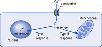
The mechanisms by which extracellular signals communicate their message across the neuron membrane to alter the protein production are discussed in Chapter 3. Here it will suffice to say that special transmission proteins called immediate early genes (IEG) are activated by a variety of second messenger systems in the neuron in response to membrane stimulus (Mitchell & Tjian 1989). Type 1 IEG responses are specific for the genes in the nucleus of the neuron and type 2 IEG responses are specific for mitochondrial DNA (Fig. 1.4).

Figure 1.4 Immediate early genes responses type I and type II. Following receptor activation the entry of calcium (Ca+) ions into the neuron activate both type I and type II response cascades. The type I cascade involves the activation of third-order messengers that modulate the activation or inhibition of DNA in the nucleus of the neuron. The type II cascade involves the activation of third-order messengers that modulate activation or inhibition of the mitochondrial DNA of the neuron.
Proteins have a multitude of functions in the neuron, some of which include cytoskeletal structure formation of microtubules and microfilaments, neurotransmitter production, intracellular signalling, formation of membrane receptors, formation of membrane channels, structural support of membranes, and enzyme production.
If the cell does not produce enough protein the cell cannot perform the necessary functions to the extent required for optimal performance and/or to sustain its very life.
In situations where the neuron has not had adequate supplies of oxygen, nutrients, or stimulus, the manufacturing of protein is down-regulated. This process of degeneration of function is referred to as transneural degeneration.
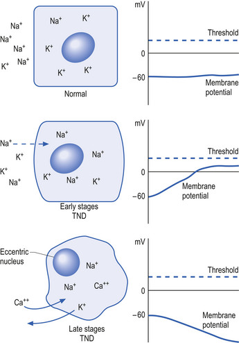
Initially, the neuron response to this down-regulation is to increase its sensitivity to stimulus so that less stimulus is required to stimulate protein production. This essentially means that the neuron alters its membrane potential so that it is closer to its threshold potential; in other words, it becomes more depolarised and becomes more irritable to any stimulus it may receive.
After a period of time if the neuron does not receive the deficient component in sufficient amounts, it can no longer sustain its state of depolarisation and starts to drastically downgrade the production of protein as a last ditch effort to conserve energy and maintain survival. At this stage, the neuron will still respond to stimulus but only for short periods as it consumes its available protein and ATP stores very quickly. In this state the neuron is vulnerable to overstimulation that may further exhaust and damage the neuron (Fig. 1.5).

Figure 1.5 The progression of transneural degeneration in a neuron. (Top) A normal healthy neuron with a normal distribution of sodium (Na+) and potassium (K+) ions across its membrane, resulting in a normal resting membrane potential. Note the central nucleus. (Middle) The early stages of transneural degeneration. In this stage, the Na+ ion concentration in the cell increases because of loss of Na+/K+ pump activity and alterations in membrane permeability, resulting in a membrane potential more positive and closer to the threshold of firing of the neuron. A neuron in this state will fire action potentials when normally inadequate stimuli are received. This inappropriate firing is called physiological irritability. The neuron will only be able to maintain the frequency of firing for short periods because of the lack of sufficient enzymes and ATP supplies before it fatigues and fails to produce action potentials. (Bottom) The neuron in the late stages of transneural degeneration. Note the eccentric nucleus, which can no longer be maintained by the degraded state of the microfilaments and microtubules. The membrane has lost its ability to segregate ions, and calcium ions have entered the neuron in high concentrations, which will eventually result in cell death. The resting membrane potential has shifted away from firing threshold, resulting in the neuron requiring excessive stimulus in order to fire an action potential.
The process of transneural degeneration may be one approach that determines the survival or death of neurons during embryological development where it has become quite clear that neurons that do not receive adequate stimulus do not usually survive (see Chapter 2).
Frequency of firing of a neuron or neuron system
The frequency of firing (FOF) of a neuron is quite simply the number of action potentials that it generates over a defined period of time. As a rule, the FOF is an important indicator of the central integrative state of a neuron. Neurons with high and regular FOF usually maintain high levels of energy and protein production that maintains the neuron in a good state of ‘health’. One exception to this rule is a neuron that is in the early stages of transneural degeneration, in which case it will produce high FOF but only for short durations.
Time to activation of a neuron or neuron system
The time to activation (TTA) of a neuron is a measure of the time from which the neuron receives a stimulus to the time that an activation response can be detected. Obviously, in clinical practice the response of individual neurons cannot be measured but the response of neuron systems such as the pupil response to light can be. As a rule, the TTA will be less (faster) in situations where the neuron system has maintained a high level of integration and activity and greater (slower) in situations where the neuron has not maintained a high level of integration and activity or is in the late stages of transneural degeneration. Again, an exception to this rule can occur in situations where the neuron system is in the early stages of transneural degeneration and is irritable to stimulus and responds quickly. This response will be of short duration and cannot be maintained for more than a short period of time.
Time to fatigue in a neuron or neuron system
The time to fatigue (TTF) in a neuron is the length of time that a response can be maintained during a continuous stimulus to the neuron. The TTF effectively measures the ability of the neuron to sustain activation under continuous stimuli, which is a good indicator of the ATP and protein stores contained in the neuron. This in turn is a good indication of the state of health of the neuron. The TTF will be longer in neurons that have maintained high levels of integration and stimulus and shorter in neurons that have not. TTF can be very useful in determining whether a fast time to response (TTR) is due to a highly integrated neuron system or a neuron system that is in the early stages of transneural degeneration.
For example, in clinical practice we can compare the individual responses of two pupils to light. If both pupils respond very quickly to light stimulus (fast TTR), and they both maintain pupil contraction for 3–4 seconds (long TTF), this is a good indication that both neuronal circuits are in a good state of health. If, however, both pupils respond quickly (fast TTR) but the right pupil immediately dilates despite the continued presence of the light stimulus (short TTF), this may be an indication that the right neuronal system involved in pupil constriction may be in an early state of transneural degeneration and more detailed examination is necessary.
Diaschisis
Diaschisis refers to the process of degeneration of a downstream neuronal system in response to a decrease in stimulus from an upstream neuronal system.
This reemphasises the point that neuronal systems do not exist in isolation but are involved in highly complicated and interactive networks. Interference or disruption in one part of the network can impact other parts of the network.
For example, injury or disease affecting the cerebellum invariably also affects the activity of the contralateral thalamus and cortex.
Constant and non-constant neural pathways
All multimodal integrated neuronal systems need to receive input from a constant stimulus pathway as well as appropriate oxygen and nutrient supply in order to maintain a healthy CIS.
Constant stimulus pathways are neural receptive systems that supply constant input into the neuraxis that are integrated throughout all multimodal systems to provide the stimulus necessary for the development and maintenance of the systems. Examples of constant stimulus pathways include receptors that detect the effects of gravity or constant motion, namely the joint and muscle position receptors of joint capsules and muscle spindles of the midline or axial structures including the ribs and spinal column. Certain aspects of the vestibulocerebellar system receive constant input and are constantly active. Several neural systems contain groups of neurons that exhibit innate pacemaker depolarisation mechanisms such as cardiac pacemaker cells, certain thalamic neurons, and selective neurons of the basal ganglia.
All other receptor systems are non-constant in nature, which means they are activated in bursts of activity that are not constantly maintained.

Stay updated, free articles. Join our Telegram channel

Full access? Get Clinical Tree

