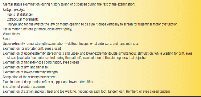
The complete neurologic examination can be a complex and arduous undertaking. In fact, few neurologists do a truly complete exam on every patient. As with the general physical examination, the history focuses the neurologic examination so that certain aspects are emphasized in a given clinical situation. The exam done on a typical patient with headache is not the same as that done on a patient with low back pain, or dementia, or cerebrovascular disease. The examination should also be adapted for the circumstances. If the patient is in pain or feels apprehensive, it may initially focus on the area of complaint, followed later by a more thorough assessment. Only a brief examination may be possible for unstable or severely ill persons until their condition stabilizes. With comatose, combative, or uncooperative patients, a compulsively complete examination is an impossibility. However, in each of these situations, at least some maneuvers are employed to screen for neurologic dysfunction that is not necessarily suggested by the history. A rapid screening or mini neurologic examination may initially be adequate for persons with minor or intermittent symptoms. Every patient does not require every conceivable test, but all require a screening examination. The findings on such a screening examination determine the emphasis of a more searching subsequent examination. There are a number of ways to perform a screening examination. Table 5.2 details such an abbreviated examination from previous editions of this book.
TABLE 5.2 Components of a Screening Initial Neurologic Examination (Abnormalities or Specific Symptoms Should Lead to More Complete Evaluations)

There are two basic ways to do a traditional neurologic examination—regional and systemic. A system approach evaluates the motor system, then the sensory system, and so on. A regional approach evaluates all the systems in a given region, such as the upper extremities and then the lower extremities. The screening exam outlined in Table 5.3 is an amalgam of the regional and system approaches geared for speed and efficiency. The concept is an examination that requires the nervous system to perform at a high level, relying heavily on sensitive signs, especially the flawless execution of complex functions. If the nervous system can perform a complex task perfectly, it is very unlikely there is significant pathology present, and going through a more extensive evaluation is not likely to prove productive. A neurologic examination that assesses complex functions and seeks signs that are sensitive indicators of pathology is efficient and not overly time consuming.
TABLE 5.3 Steps in a Screening Neurologic Examination

Modified from: Campbell WW, Pridgeon RP. Practical Primer of Clinical Neurology. Philadelphia: Lippincott Williams & Wilkins, 2002.
Educators have proposed a third type of exam, especially for teaching: the hypothesis-driven examination. This approach evolves naturally with experience but has not been used previously in teaching the exam. Teaching a hypothesis-driven neurologic exam evolved from a similar approach to the general physical exam. Examination maneuvers were focused by the history. Using the hypothesis-driven approach produced greater sensitivity but lower specificity, and was performed in less time. Learning to develop a hypothesis from the history and how to test it is of course a paramount challenge in neurology.
The examination begins with taking the medical history, which serves as a fair barometer of the mental status. Patients who can relate a logical, coherent, pertinent, and sensible narrative of their problem will seldom have abnormalities on more formal bedside mental status testing. On the other hand, a rambling, disjointed, incomplete history may be a clue to the presence of some cognitive impairment, even though there is no direct complaint of thinking or memory problems from the patient or the family. Similarly, psychiatric disease is sometimes betrayed by the patient’s demeanor and style of history giving. If there is any suggestion of abnormality from the interaction with the patient during the history-taking phase of the encounter, then a more detailed mental status examination should be carried out. Other reasons to do a formal mental status examination are discussed in Chapter 8. Simple observation is often useful. The patient’s gait, voice, mannerisms, ability to dress and undress, and even handshake (grip myotonia) may suggest the diagnosis.
The screening examination detailed in Table 5.3 continues by doing everything that requires use of a penlight. Begin by noting the position of the eyelids and the width of the palpebral fissures bilaterally. Check the pupils for light reaction with the patient fixing at distance. If the pupillary light reaction is normal and equal in both eyes, checking the pupillary near reaction is not necessary. Continue by assessing extraocular movements in the six cardinal positions of gaze, having the patient follow the penlight. Be sure the patient has no diplopia or limitation of movement and that ocular pursuit movements are smooth and fluid. With the eyes in primary and eccentric positions, look for any nystagmus. The eye examination is discussed in more detail in Chapter 14
Stay updated, free articles. Join our Telegram channel

Full access? Get Clinical Tree


