Chapter 3 Gross Anatomy and General Organization of the Central Nervous System
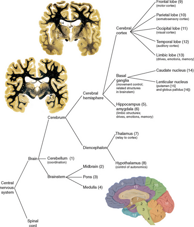
The human central nervous system (CNS) is composed of the brain and spinal cord. This chapter briefly discusses the major surface and internal structures of the brain (summarized in Fig. 3-25) and, together with the next six chapters, lays the groundwork for the more detailed discussion of the functional anatomy of the CNS in subsequent chapters.
The Long Axis of the CNS Bends at the Cephalic Flexure
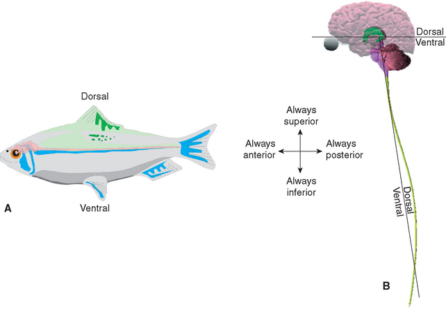
Before considering the parts of the brain in more detail, it is helpful to discuss some terms used for planes and directions in the nervous system. The sagittal plane divides the brain into two symmetrical halves. Parasagittal planes are those parallel to the sagittal plane. Coronal planes (also called frontal planes) are parallel to the long axis of the body and perpendicular to the sagittal plane (e.g., a vertical plane passing through both ears). Axial planes (also called transverse or horizontal planes) are perpendicular to the long axis of the body. These terms are fairly straightforward and have the same meaning with respect to any part of the nervous system. However, directional terms such as anterior, dorsal, and rostral change their meanings relative to one another in different parts of the nervous system. The reason for this, as indicated in Figure 3-1, is that walking upright necessitates a bend of about 80 degrees in going from the long axis of the spinal cord and brainstem to the long axis of the cerebrum. This bend is a consequence of the cephalic flexure, which appears early in the embryological development of the nervous system (see Fig. 2-8) and persists in the mature brain. Dorsal-ventral terminology ignores this bend, as though we had a linear CNS and walked around on all fours like most other vertebrates, so that the directional meaning of dorsal changes by 80 degrees at the midbrain-diencephalon junction. The terms anterior and superior, in contrast, retain a constant meaning relative to the normal upright orientation of the body as a whole. This means, for example, that the ventral surface of the spinal cord is also its anterior surface, but the ventral surface of the diencephalon is its inferior surface. Rostral-caudal terminology may cause additional confusion. Anatomically, rostral means “toward the nose.” However, it also has a functional connotation (implying “toward the telencephalon”), so that the posterior end of the cerebral hemispheres could be considered rostral to all parts of the diencephalon. Use of anterior-posterior and superior-inferior (or dorsal-ventral) terminology in reference to the cerebrum avoids any ambiguity.
Hemisecting a Brain Reveals Parts of the Diencephalon, Brainstem, and Ventricular System
The cerebral hemispheres conceal most of the rest of an intact brain (Fig. 3-2A). Hemisection reveals many parts of the diencephalon, brainstem, and cerebellum, and additional features of the cerebral hemispheres as well (Fig. 3-2B). The cephalic flexure is visible at the junction between the brainstem and the diencephalon. The brainstem itself is subdivided into the midbrain, which is continuous with the diencephalon; the pons; and the medulla, which is continuous with the spinal cord. The two cerebral hemispheres are joined by a huge fiber bundle, the corpus callosum, which has an enlarged and rounded posterior splenium, a body, and an anterior, curved genu that tapers gently into a ventrally directed rostrum, which merges into the lamina terminalis (where development of the corpus callosum started).
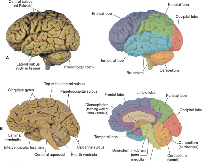
The nervous system develops embryologically from a neuroectodermal tube; the cavity of the tube persists in adults as a system of ventricles (see Fig. 5-1), part of which is apparent in the sagittal plane (Fig. 3-2B). Portions of the medial surfaces of the diencephalon form the walls of the narrow, slitlike third ventricle, which opens into the large lateral ventricle of each cerebral hemisphere through an interventricular foramen (foramen of Monro). Posteriorly, the third ventricle is continuous with a narrow channel through the midbrain, the cerebral aqueduct (aqueduct of Sylvius). The aqueduct in turn is continuous with the fourth ventricle of the pons and medulla, and the fourth ventricle is continuous with the microscopically tiny central canal of the caudal medulla and the spinal cord.
Humans, Relative to Other Animals, Have Large Brains
One impressive feature of the human brain is its size, and our distinctively human mental capacities are commonly attributed to this. The human brain weighs about 400 g at birth, and this weight triples during the first 3 years of life (resulting from the addition of myelin and the growth of neuronal processes rather than the addition of neurons). The rate of growth then slows, and the maximum brain weight of around 1400 g (Table 3-1) is reached at about age 11. This weight holds steady until about age 50, when a slow decline sets in (Fig. 3-3). The weight of 1400 g is only an average; brain weights for normal individuals range from 1100 g (or less) to around 1700 g. This large range is surprising, and its significance is not well understood; there is only a modest correlation between brain size and mental ability.
Table 3-1 Approximate Average Volumes of Intracranial Contents
| Volume (cm3) | |
|---|---|
| Brain | 1365 |
| Gray matter | 695 |
| White matter | 670 |
| CSF | 180 |
| Cerebrum | 1200 |
| Basal ganglia* | 8 |
| Cerebellum | 135 |
| Brainstem | 30 |
Based on Anastasi et al (2006), Kruggel (2006), and Makris et al (2003).
* Caudate nucleus, putamen, and globus pallidus.
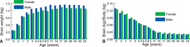
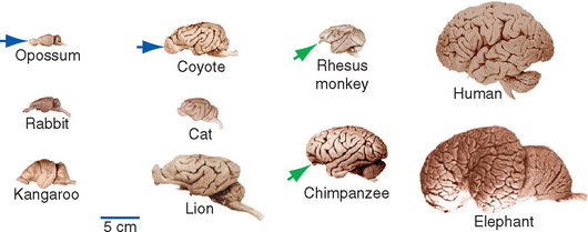
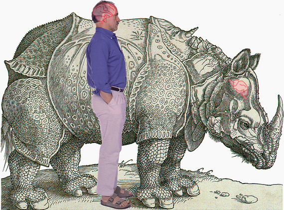
Part of the reason for the large human brain is simply a reflection of body size: big animals tend to have big brains (Fig. 3-4). Elephants, for example, have 5000-g brains. Similarly, the size difference between the bodies of human males and females explains, at least in part, the fact that male brains are slightly larger than female brains (Fig. 3-3). However, this is not the whole story; many animals that are larger than humans nevertheless have smaller brains (Fig. 3-5). Overall, then, relative to body size, humans have larger brains than most other animals. It is tempting to attribute our mental abilities to our relatively large brains, but this is an oversimplification. Relative to body size, dolphins, some small primates and rodents, and even some fish have larger brains than we do. The key differences between human brains and those of other animals appear to be more complex neuronal interconnections and a selective increase in the size of certain areas of the cerebral cortex thought to be involved in higher functions (see Fig. 22-12).
Named Sulci and Gyri Cover the Cerebral Surface
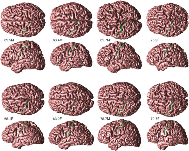
A striking aspect of human cerebral hemispheres is the degree to which their surface is folded and convoluted. Each ridge is called a gyrus, and each groove between ridges is called a sulcus; particularly deep sulci are often called fissures. This folding into gyri and sulci is a mechanism for increasing the total cortical area; each of us has about 2.5 ft2 of cortex, two thirds of which is hidden from view in the walls of sulci. The appearance of different gyri and sulci varies considerably from one brain to another (Fig. 3-6), to the extent that they may not even be continuous structures (e.g., a particular gyrus may be transected by one or more sulci). Major features, however, are reasonably constant.
In the discussion that follows, the principal surface features of the hemispheres are described, with some broad generalizations regarding the function of various cortical areas. These functional descriptions are highly oversimplified and are offered primarily for purposes of initial orientation. Cortical function is discussed in more detail in Chapter 22.
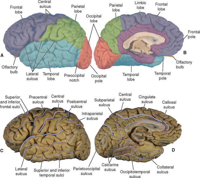
Each Cerebral Hemisphere Includes a Frontal, Parietal, Occipital, Temporal, and Limbic Lobe
Four prominent sulci—the central sulcus, the lateral sulcus, the parietooccipital sulcus, and the cingulate sulcus—together with the preoccipital notch and parts of a few other sulci are used to divide each cerebral hemisphere into five lobes (Fig. 3-7):
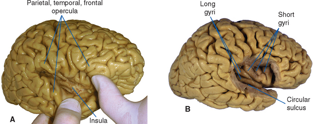
An additional area of cerebral cortex not usually included in any of the five lobes discussed above lies buried in the depths of the lateral sulcus, concealed from view by portions of the frontal, parietal, and temporal lobes. This cortex, called the insula, overlies the site where the telencephalon and diencephalon fuse during embryological development (see Chapter 2). It can be revealed by prying open the lateral sulcus or by removing the overlying portions of other lobes (Fig. 3-8). The portion of a given lobe overlying the insula is called an operculum (Latin for “lid”); there are frontal, parietal, and temporal opercula. The circular sulcus outlines the insula and marks its borders with the opercular areas of cortex.
The Frontal Lobe Contains Motor Areas
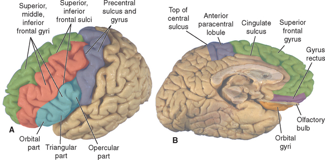
Four gyri make up the lateral surface of the frontal lobe (Fig. 3-9). The precentral gyrus is anterior to the central sulcus and parallel to it, extending to the precentral sulcus. The superior, middle, and inferior frontal gyri are oriented parallel to one another and roughly perpendicular to the precentral gyrus. The superior frontal gyrus continues onto the medial surface of the hemisphere as far as the cingulate sulcus. The inferior frontal gyrus is visibly divided into three parts: (1) the orbital part, which is most anterior and is continuous with the inferior (orbital) surface of the frontal lobe; (2) the opercular part, which is most posterior and forms a portion of the frontal operculum; and (3) the wedge-shaped triangular part, which lies between the other two. The inferior, or orbital, surface of the frontal lobe is mostly occupied by a group of gyri of somewhat variable appearance that are usually referred to collectively as orbital gyri or orbitofrontal cortex. The only consistently named gyrus on this surface is the gyrus rectus (Greek for “straight gyrus”), which is most medial and extends onto the medial surface of the hemisphere. Between the gyrus rectus and the orbital gyri is the olfactory sulcus, which contains the olfactory bulb and tract. The medial surface of the lobe contains extensions of the superior frontal gyrus, precentral gyrus, and gyrus rectus; certain small cortical areas near the rostrum of the corpus callosum are part of the limbic lobe.
The frontal lobe contains four general functional areas:
The Parietal Lobe Contains Somatosensory Areas
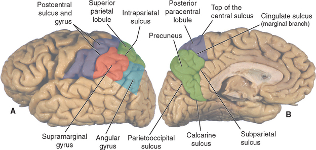
The lateral surface of the parietal lobe is divided into three areas: the postcentral gyrus and the superior and inferior parietal lobules (Fig. 3-10). The postcentral gyrus is posterior to the central sulcus and parallel to it, extending to the postcentral sulcus. The intraparietal sulcus runs posteriorly from the postcentral sulcus toward the occipital lobe, separating the superior and inferior parietal lobules. The inferior parietal lobule in turn is composed of the supramarginal gyrus, which caps the upturned end of the lateral sulcus, and the angular gyrus, which similarly caps the superior temporal sulcus. The angular gyrus is typically broken up by small sulci and may overlap the supramarginal gyrus. The medial surface of the parietal lobe contains the medial extension of the postcentral gyrus and is completed by an area called the precuneus, which is bounded by the subparietal and calcarine sulci, the parietooccipital sulcus, and the marginal branch of the cingulate sulcus. The extensions of the precentral and postcentral gyri onto the medial surface of the hemisphere are sometimes referred to together as the paracentral lobule, which is partly in the frontal lobe and partly in the parietal lobe.
< div class='tao-gold-member'>


Stay updated, free articles. Join our Telegram channel

Full access? Get Clinical Tree


