Chapter 1 History of Sleep Physiology and Medicine
Abstract
Interest in sleep and dreams has existed since the dawn of history. Perhaps only love and human conflict have received more attention from poets and writers. Some of the world’s greatest thinkers, such as Aristotle, Hippocrates, Freud, and Pavlov, have attempted to explain the physiologic and psychological bases of sleep and dreaming. However, it is not the purpose of this chapter to present a scholarly review across the ages about prehistoric, biblical, and Elizabethan thoughts and concerns regarding sleep or the history of man’s enthrallment with dreams and nightmares. This has been reviewed by others.1 What is emphasized here for the benefit of the student and the practitioner is the evolution of the key concepts that define and differentiate sleep research and sleep medicine, crucial discoveries and developments in the formative years of the field, and those principles and practices that have stood the test of time.
Sleep as a Passive State
“Sleep is the intermediate state between wakefulness and death; wakefulness being regarded as the active state of all the animal and intellectual functions, and death as that of their total suspension.”2
The passive to active historical dichotomy is also given great weight by the modern investigator J. Allan Hobson.3 In the first sentence of his book, Sleep, published in 1989, he stated that “more has been learned about sleep in the past 60 years than in the preceding 6,000.” He went on, “In this short period of time, researchers have discovered that sleep is a dynamic behavior. Not simply the absence of waking, sleep is a special activity of the brain, controlled by elaborate and precise mechanisms.”
The hypnotoxin theory reached its zenith in 1907 when French physiologists, Legendre and Pieron,4 did experiments showing that blood serum from sleep-deprived dogs could induce sleep in dogs that were not sleep deprived. The notion of a toxin causing the brain to sleep has gradually given way to the notion that there are a number of endogenous “sleep factors” that actively induce sleep by specific mechanisms.
In the 1920s, the University of Chicago physiologist Nathaniel Kleitman carried out a series of sleep-deprivation studies and made the simple but brilliant observation that individuals who stayed up all night were generally less sleepy and impaired the next morning than in the middle of their sleepless night. Kleitman argued that this observation was incompatible with the notion of a continual buildup of a hypnotoxin in the brain or blood. In addition, he felt that humans were about as impaired as they would get, that is, very impaired, after about 60 hours of wakefulness, and that longer periods of sleep deprivation would produce little additional change. In the 1939 (first) edition of his comprehensive landmark monograph “Sleep and Wakefulness” Kleitman5 summed up by saying, “It is perhaps not sleep that needs to be explained, but wakefulness, and indeed, there may be different kinds of wakefulness at different stages of phylogenetic and ontogenetic development. In spite of sleep being frequently designated as an instinct, or global reaction, an actively initiated process, by excitation or inhibition of cortical or subcortical structures, there is not a single fact about sleep that cannot be equally well interpreted as a let down of the waking activity.”
The Electrical Activity of the Brain
However, it was not until 1928 when the German psychiatrist Hans Berger6 recorded electrical activity of the human brain and clearly demonstrated differences in these rhythms when subjects were awake or asleep that a real scientific interest commenced. Berger correctly inferred that the signals he recorded, which he called “electroencephalograms,” were of brain origin. For the first time, the presence of sleep could be conclusively established without disturbing the sleeper, and, more important, sleep could be continuously and quantitatively measured without disturbing the sleeper.
All the major elements of sleep brain wave patterns were described by Harvey, Hobart, Davis, and others7–9 at Harvard University in a series of extraordinary papers published in 1937, 1938, and 1939. Blake, Gerard, and Kleitman10,11 added to this from their studies at the University of Chicago. On the human electroencephalogram (EEG), sleep was characterized by high-amplitude slow waves and spindles, whereas wakefulness was characterized by low-amplitude waves and alpha rhythm. The image of the sleeping brain completely “turned off” gave way to the image of the sleeping brain engaged in slow, synchronized, “idling” neuronal activity. Although it was not widely recognized at the time, these studies were some of the most critical turning points in sleep research. Indeed, Hobson3 dated the turning point of sleep research to 1928, when Berger began his work on the human EEG. Used today in much the same way as they were in the 1930s, brain wave recordings with paper and ink, or more recently on computer screens, have been extraordinarily important to sleep research and sleep medicine.
The 1930s also saw one series of investigations that seemed to establish conclusively both the passive theory of sleep and the notion that it occurred in response to reduction of stimulation and activity. These were the investigations of Frederick Bremer,12,13 reported in 1935 and 1936. These investigations were made possible by the aforementioned development of electroencephalography. Bremer studied brain wave patterns in two cat preparations. One, which Bremer called encéphale isolé, was made by cutting a section through the lower part of the medulla. The other, cerveau isolé, was made by cutting the midbrain just behind the origin of the oculomotor nerves. The first preparation permitted the study of cortical electrical rhythms under the influence of olfactory, visual, auditory, vestibular, and musculocutaneous impulses; in the second preparation, the field was narrowed almost entirely to the influence of olfactory and visual impulses.
The Ascending Reticular System
After World War II, insulated, implantable electrodes were developed, and sleep research on animals began in earnest. In 1949, one of the most important and influential studies dealing with sleep and wakefulness was published: Moruzzi and Magoun’s classic paper “Brain Stem Reticular Formation and Activation of the EEG.”14 These authors concluded that “transitions from sleep to wakefulness or from the less extreme states of relaxation and drowsiness to alertness and attention are all characterized by an apparent breaking up of the synchronization of discharge of the elements of the cerebral cortex, an alteration marked in the EEG by the replacement of high voltage, slow waves with low-voltage fast activity” (p. 455).
The demonstration by Starzl and coworkers15 that sensory collaterals discharge into the reticular formation suggested that a mechanism was present by which sensory stimulation could be transduced into prolonged activation of the brain and sustained wakefulness. By attributing an amplifying and maintaining role to the brainstem core and the conceptual ascending reticular activating system, it was possible to account for the fact that wakefulness outlasts, or is occasionally maintained in the absence of, sensory stimulation.
Chronic lesions in the brainstem reticular formation produced persisting slow waves in the EEG and immobility. The usual animal for this research was the cat because excellent stereotaxic coordinates of brain structures had become available in this model.16 These findings appeared to confirm and extend Bremer’s observations. The theory of the reticular activating system was an anatomically based passive theory of sleep or an active theory of wakefulness. Figure 1-1 is from the published proceedings of a symposium entitled Brain Mechanisms and Consciousness, which published in 1954 and is probably the first genuine neuroscience bestseller.17 Horace Magoun had extended his studies to the monkey, and the illustration represents the full flowering of the ascending reticular activating system theory.
Early Observations of Sleep Pathology
Obstructive sleep apnea syndrome (OSAS), which may be called the leading sleep disorder of the 20th century, was described in 1836, not by a clinician but by the novelist Charles Dickens. In a series of papers entitled the “Posthumous Papers of the Pickwick Club,” Dickens described Joe, a boy who was obese and always excessively sleepy. Joe, a loud snorer, was called Young Dropsy, possibly as a result of having right-sided heart failure. Meir Kryger18 and Peretz Lavie19,20 published scholarly accounts of many early references to snoring and conditions that were most certainly manifestations of OSAS. Professor Pierre Passouant21 provided an account of the life of Gélineau and his landmark description of the narcolepsy syndrome.
Chronobiology
Most, but not all, sleep specialists share the opinion that what has been called chronobiology or the study of biologic rhythms is a legitimate part of sleep research and sleep medicine. The 24-hour rhythms in the activities of plants and animals have been recognized for centuries. These biologic 24-hour rhythms were quite reasonably assumed to be a direct consequence of the periodic environmental fluctuation of light and darkness. However, in 1729, Jean Jacques d’Ortous de Mairan described a heliotrope plant that opened its leaves during the day even after de Mairan had moved the plant so that sunlight could not reach it. The plant opened its leaves during the day and folded them for the entire night even though the environment was constant. This was the first demonstration of the persistence of circadian rhythms in the absence of environmental time cues. Figure 1-2, which represents de Mairan’s original experiment, is reproduced from The Clocks That Time Us by Moore-Ede and colleagues.22
The Discovery of REM Sleep
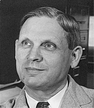
The characterization of rapid eye movement (REM) sleep as a discrete organismic state should be distinguished from the discovery that rapid eye movements occur during sleep. The historical threads of the discovery of rapid eye movements can be identified. Nathaniel Kleitman (Fig. 1-3, Video 1-1![]() ), a professor of physiology at the University of Chicago, had long been interested in cycles of activity and inactivity in infants and in the possibility that this cycle ensured that an infant would have an opportunity to respond to hunger. He postulated that the times infants awakened to nurse on a self-demand schedule would be integral multiples of a basic rest-activity cycle. The second thread was Kleitman’s interest in eye motility as a possible measure of “depth” of sleep. The reasoning for this was that eye movements had a much greater cortical representation than did almost any other observable motor activity, and that slow, rolling, or pendular eye movements had been described at the onset of sleep with a gradual slowing and disappearance as sleep “deepened.”23
), a professor of physiology at the University of Chicago, had long been interested in cycles of activity and inactivity in infants and in the possibility that this cycle ensured that an infant would have an opportunity to respond to hunger. He postulated that the times infants awakened to nurse on a self-demand schedule would be integral multiples of a basic rest-activity cycle. The second thread was Kleitman’s interest in eye motility as a possible measure of “depth” of sleep. The reasoning for this was that eye movements had a much greater cortical representation than did almost any other observable motor activity, and that slow, rolling, or pendular eye movements had been described at the onset of sleep with a gradual slowing and disappearance as sleep “deepened.”23

Figure 1-3 Nathaniel Kleitman (circa 1938), Professor of Physiology, University of Chicago, School of Medicine.
At this point, Aserinsky and Kleitman made two assumptions:
Aserinsky and Kleitman initiated a small series of awakenings, both when rapid eye movements were present and when rapid eye movements were not present, for the purpose of eliciting dream recall. They did not apply sophisticated methods of dream content analysis, but the descriptions of dream content from the two conditions were generally quite different with REM awakenings yielding vivid complex stories and non-REM (NREM) awakenings often yielding nothing at all or very sparse accounts. This made it possible to conclude that rapid eye movements were associated with dreaming. This was, indeed, a breakthrough in sleep research.24,25
All-Night Sleep Recordings and the Basic Sleep Cycle
However, motivated by the desire to expand and quantify the description of rapid eye movements, then graduate student William Dement and Kleitman26 did just this over a total of 126 nights with 33 subjects and, by means of a simplified categorization of EEG patterns, scored the paper recordings in their entirety. When they examined these 126 records, they found that there was a predictable sequence of patterns over the course of the night, such as had been hinted at by Aserinsky’s study but entirely overlooked in all previous EEG studies of sleep. Although this sequence of regular variations has now been observed tens of thousands of times in hundreds of laboratories, the original description remains essentially unchanged.
The usual sequence was that after the onset of sleep, the EEG progressed fairly rapidly to stage 4, which persisted for varying amounts of time, generally about 30 minutes, and then a “lightening” took place. Whereas the progression from wakefulness to stage 4 at the beginning of the cycle was almost invariable through a continuum of change, the lightening was usually abrupt and coincident with a body movement or series of body movements. After the termination of stage 4, there was generally a short period of stage 2 or stage 3 which gave way to stage 1 and rapid eye movements. When the first eye movement period ended, the EEG again progressed through a continuum of change to stage 3 or 4, which persisted for a time and then lightened, often abruptly, with body movement to stage 2, which again gave way to stage 1 and the second rapid eye movement period (see p. 679 of Dement and Kleitman26).
Stay updated, free articles. Join our Telegram channel

Full access? Get Clinical Tree


