CHAPTER 127 Intracranial Ependymomas in Adults
Intracranial ependymomas in adults are relatively rare brain tumors of neuroectodermal origin that account for 2% to 6% of all intracranial neoplasms and occur equally in males and females.1–6 These lesions are more common in children, in whom they account for 10% of brain tumors.7 Supratentorial ependymomas occur more frequently in adults, whereas infratentorial tumors are more common in the pediatric population (Table 127-1).7–15 Surgical resection is regarded as the standard treatment of these tumors, with a goal of gross total resection when safely feasible. The optimal therapeutic management of intracranial ependymomas in adults remains controversial because of the rarity of these lesions in this population and the limited number of studies pertaining to adults. The majority of studies on intracranial ependymomas have been conducted in the pediatric population. The low incidence of these tumors in adult patients has led to many published reports that are retrospective, included both intracranial and spinal ependymomas in some studies, combined data from children and adults in some series, and covered long periods of treatment, which confounds the interpretation of results because of changes in histologic grading, diagnosis, and therapy, all of which have contributed to the lack of ability to reach a consensus in optimally managing these lesions in adults. However, adjuvant postoperative radiotherapy (RT) is typically considered to play an important role in treatment, particularly in the care of patients with high-grade ependymomas, whereas its role in the management of low-grade tumors continues to remain controversial in adults.4,6,16,17 Furthermore, a role for adjuvant chemotherapy has not been adequately defined.
TABLE 127-1 Comparison of Intracranial Ependymomas in Adults and Children
| ADULT | PEDIATRIC | |
|---|---|---|
| Prevalence | 2%-6% of intracranial neoplasms | 10% of intracranial neoplasms |
| Location | Predominantly supratentorial | Predominantly infratentorial |
| Cerebrospinal fluid seeding | Less common | More common |
| Clinical studies | Paucity because of low incidence | Several because of higher incidence |
| 5-Year survival rate | 55%-90% | 40%-65% |
Although prospective studies are needed in adult patients harboring intracranial ependymomas, reports have found a trend for better survival in adults than in children.12,18–21
Pathology
Ependymal tumors are presumed to be derivatives of the neuroectodermal cell lineage that give rise to the ependymal cells lining the choroid plexus and white matter adjacent to the angulated ventricles, especially the regions adjacent to the trigone of the lateral ventricle and the foramen of Luschka, as well as the central canal of the spinal cord for tumors affecting this part of the neuraxis.1 These ependymal cells undergo neoplastic transformation leading to ependymomas. Ependymomas located in the brain parenchyma are thought to occur as a result of fetal ependymal cell rests remaining within the parenchyma during embryogenesis.22,23 It has been estimated that approximately 50% of supratentorial ependymomas originate in the brain parenchyma and the remaining tumors are derived from the lateral ventricles, with very few originating from the third ventricle.23 Although rare, supratentorial intracortical and intracranial ectopic ependymomas have been reported.24–34 Intracranial ectopic ependymomas occur where ependymal cells are typically absent and have been reported to involve the neurohypophysis, cranial nerve V, sella turcica, posterior fossa, falx, and cavernous sinus.27–34
The World Health Organization (WHO) has classified ependymal tumors into three grades according to cellular derivatives or degree of anaplasia: (1) myxopapillary ependymoma and subependymoma (WHO grade I), (2) ependymoma (WHO grade II), and (3) anaplastic ependymoma (WHO grade III).35–39 Gross inspection of ependymomas reveals them to be solid, well-delineated, dark, gray-red lesions. Myxopapillary ependymomas occur almost exclusively in the conus, cauda equina, and filum terminale regions, and subependymomas are benign, slow-growing tumors whose diagnosis portends a favorable prognosis. Histopathologic examination of ependymomas reveals that they are composed of uniform cuboidal and astrocyte-like fibrillary cells arranged into linear tubules in which the cells surround a central lumen and resemble ependymal epithelia. These histologic findings are known as rosettes. Immunostaining for glial fibrillary acidic protein (GFAP) is typically positive, and these GFAP-positive processes can be found aligned around blood vessels and are known as perivascular pseudorosettes.40 The rosettes and perivascular pseudorosettes are key histologic features of ependymomas.
Moreover, low-grade ependymomas, which are the prevalent histologic type in the adult population, are further stratified into four subtypes: (1) cellular, (2) papillary, (3) clear cell, and (4) tanycytic. Cellular ependymomas are hypercellular with rare or absent mitoses. Papillary ependymomas, which have a tubulovillous arrangement, have a very low incidence, and immunostaining is positive for GFAP and vimentin. The clear cell subtype is characterized by clear cytoplasm and can resemble oligodendrogliomas, central neurocytomas, hemangioblastomas, or metastatic clear cell carcinomas.35,41 Therefore, careful microscopic examination of the specimen is necessary. Tanycytic ependymomas may appear histologically similar to astrocytomas given that their perivascular pseudorosettes are less obvious and rosettes are usually absent. Microscopic examination demonstrates elongated paraventricular glial cells with cytoplasmic processes that extend to the ependymal surface.42
Anaplastic ependymomas are characterized by histologic features such as hypercellularity, frequent mitotic figures, pseudopalisading necrosis, vascular proliferation, and cellular and nuclear pleomorphism.43 Rosettes are usually absent or rare, perivascular pseudorosettes may be poorly demarcated, and there may be a reduction in immunostaining for GFAP when compared with conventional ependymomas. The diagnosis of poorly differentiated lesions may require electron microscopic analysis if determination of the pathology is difficult. These tumors are locally invasive and have a higher propensity to spread to other areas of the neuraxis via cerebrospinal fluid (CSF) pathways.
Molecular Genetics
Molecular analysis has thus far demonstrated cytogenetic heterogeneity, although the data are limited. Aberrations involving chromosome 22 have been the most commonly implicated in ependymal tumorigenesis, including monosomy 22, various translocations, or the possible absence of tumor suppressor genes.44–47 The 22q region has been studied because it contains the neurofibromatosis type 2 (NF2) tumor suppressor gene. Patients with NF2 have an increased predilection for the development of ependymomas and meningiomas, and mutations in the NF2 gene have been detected in tumor specimens from these patients, in contrast to ependymomas observed in patients without NF2, in whom mutations in the NF2 gene have not been revealed.48–50 Studies have reported a loss of chromosome arm 22q in 50% to 60% of adult patients, and chromosome 22 has been implicated in the occurrence of familial intracranial ependymomas.46,51–60 In addition, abnormalities of chromosomes 1, 6, 7, 9, 10, 11, 12, 13, 16, 17, 19, and 20 have been associated with the development of ependymomas, although there are less data pertaining to them.61 Amplification of the gene MDM2 has been demonstrated in approximately 35% of ependymomas, but the role of its product, MDM2, has not been defined in the tumorigenesis of ependymomas.62 The development of new molecular techniques should aid in elucidation of the molecular mechanisms underlying the tumorigenesis of ependymomas.
Clinical Features
The clinical findings in patients with ependymomas are contingent on the size, location, and malignancy of the tumor. Ependymomas typically increase in size slowly and may reach a large dimension before detection.63 Anaplastic ependymomas may exhibit a more rapid onset of signs and symptoms. Although infratentorial lesions are more common in the pediatric population, they can occur in adults. Posterior fossa ependymomas may cause nausea, emesis, ataxia, hemiparesis, dizziness, nystagmus, and headaches.12,23 These symptoms are usually due to an increase in intracranial pressure as a consequence of obstructive hydrocephalus from filling of the fourth ventricle by the tumor or cerebellar compression, or both.
Supratentorial ependymomas are found more commonly in adults and tend to cause focal neurological deficits. Manifestation of the neurological deficits depends on the location of the tumor. These deficits may include motor weakness, aphasia, visual field deficits, behavioral changes, and memory impairment.8,64 Nausea, emesis, and headaches may occur as a result of an increase in intracranial pressure. Seizures may occur with extraventricular supratentorial tumors and have been reported to occur in approximately a third of patients.23 Although rare, supratentorial intracortical ependymomas have been reported in three adult patients, and all three were initially evaluated because of seizures.24,25 Parinaud’s syndrome may be observed in some patients with third ventricular ependymomas.1
Imaging
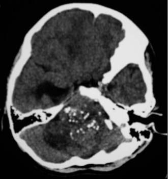
Ependymomas tend to be cystic, calcified, and well-circumscribed lesions. They may appear either isodense or hyperdense to brain parenchyma on computed tomography (CT). Administration of contrast material will typically demonstrate varying degrees of enhancement from mild to intense heterogeneous or homogeneous enhancement. CT imaging assists in the identification of any calcifications, which are present in 50% of supratentorial ependymomas and 46% of infratentorial ependymomas (Fig. 127-1).15 Imaging may be limited by artifact from the bony architecture of the posterior fossa.
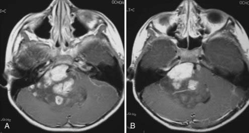
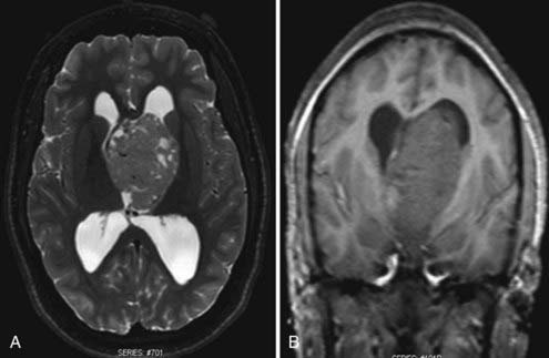
Magnetic resonance imaging (MRI) is the diagnostic modality of choice given its ability to provide greater anatomic detail. T1-weighted imaging demonstrates the tumor to be hypointense to isointense relative to white matter, whereas it is hyperintense to white matter on T2-weighted imaging. Similar to CT, administration of contrast material will typically demonstrate varying degrees of enhancement from mild to intense heterogeneous or homogeneous enhancement (Fig. 127-2). Calcium, hemosiderin, or necrosis may be demonstrated by hypointense foci on both T1- and T2-weighted images, and cystic changes are typically hyperintense on T2-weighted images (Fig. 127-3).65,66 Intratumoral hemorrhage has been observed in these lesions. Subsequently, the heterogeneity of signal characteristics on MRI could make the diagnosis of ependymoma difficult. Infratentorial ependymomas may extend from the fourth ventricle through the foramen of Luschka into the cerebellopontine cistern or downward through the foramen of Magendie into the cervical subarachnoid space.10,67,68 These characteristic findings lend support for the diagnosis of ependymoma.
Staging
If an ependymoma is suspected on imaging of the brain or confirmed with biopsy or at surgery, MRI of the spine may be performed to rule out CSF seeding given the tumor’s predilection to disseminate throughout the CSF pathways. However, no conventional staging criteria exist. Metastatic seeding has been reported to occur more frequently with WHO grade III ependymomas, posterior fossa lesions, and an inability to achieve gross total resection, as well as in the pediatric population. CSF dissemination of ependymoma has been reported to occur less frequently in the adult population.69–74
Treatment
Surgery
Surgery is the mainstay of treatment of intracranial ependymomas, and reports have regarded the extent of tumor resection to weigh heavily in patient prognosis.12,14,15,67,75,76 An attempt at gross total resection, which may increase the chance for cure or longer disease-free survival, should always be made when technically feasible. Before the advent of microsurgical techniques, some authors reported high rates of morbidity and mortality related to the performance of surgery on ependymomas, especially for lesions located in the posterior fossa. Morbidity and mortality rates have been minimized with the introduction of microsurgical techniques, intraoperative neuronavigational modalities, intraoperative ultrasound, and intraoperative neurophysiologic monitoring, which contribute to the ability to achieve gross total or near-total resection of the ependymoma when technically possible. Complete resection of supratentorial ependymomas is more likely than for those located infratentorially because of the location of key structures, such as the brainstem, cranial nerve nuclei in the floor of the fourth ventricle, and vasculature, within a small, confined area.
Supratentorial Ependymomas
Supratentorial intraventricular ependymomas can be approached through either a transcortical or interhemispheric transcallosal technique. The surgeon should have a sound knowledge base with regard to the different approaches to the ventricles. Preoperative MRI is used to assess a surgical corridor and formulate the most appropriate surgical approach based on tumor size, tumor origin, and visual angles within the ventricle. Tumor origin, in the case of ventricular tumors, refers to tumors that are primarily ventricular or parenchymal with major ventricular extension. The transcortical approaches predispose patients to cortical injury and seizures. Reports estimate the risk for postoperative seizures to range from 29% to 70% after a transcortical procedure, whereas the reported risk is 0% to 10% after a transcallosal approach.77–82 Disconnection syndrome may be observed after the interhemispheric posterior transcallosal approach.83–85
Infratentorial Ependymomas
MRI is performed within 48 hours postoperatively to assess the extent of surgical extirpation. In addition, the presence of hemorrhage, particularly in the ventricular system, is evaluated because this may exacerbate preexisting obstructive hydrocephalus. Emergency postoperative head CT should be performed for any new neurological deficits or alterations in mental status. Second-look surgery has been recommended to maximize the extent of resection in cases of unexpected residual tumor or when the decision for a staged operation has been made.86 The optimal timing of second-look surgery, however, is a subject of debate. Some authors prefer a delayed second-look surgery after adjuvant chemotherapy or RT because adjuvant therapy may reduce the tumor volume or alter its characteristics such that surgical outcomes are improved.86 Since it is unclear that adjuvant therapy is beneficial, in cases in which residual tumor is easily accessible with minimal morbidity, immediate second-look surgery might be an appropriate option to achieve radical resection of tumor.86
Stay updated, free articles. Join our Telegram channel

Full access? Get Clinical Tree


