Less Commonly Used Antiepileptic Drugs


Less Commonly Used Antiepileptic Drugs


Basim M. Uthman

Ahmad Beydoun


INTRODUCTION

Despite the availability of a large number of marketed antiepileptic drugs (AEDs), physicians use only a few to treat most patients with epilepsy. The six traditional AEDs used for epileptic seizures include phenytoin, carbamazepine, valproate, primidone, phenobarbital, and ethosuximide. In 1999, phenytoin (Dilantin) had a 42% market share of total AED prescriptions for epilepsy in the United States, proprietary (Tegretol) and generic carbamazepine had a 24% share, and divalproex sodium (Depakote) had captured 17% of the market (1). The remaining 17% of the market was held by all other products and generics. The availability of less-sedating AEDs led to a gradual decline in the use of primidone and phenobarbital in the United States. This prescribing pattern reflects the physician’s familiarity with a particular agent, its efficacy, tolerability, pharmacokinetic profile, and cost. Results of comparative trials have led to improved objectivity in the selection of the best agent for specific seizure disorders.

Over the past decade, the introduction of new AEDs—felbamate (Felbatol1), gabapentin (Neurontin), lamotrigine (Lamictal), topiramate (Topamax), tiagabine (Gabitril), zonisamide (Zonegran), oxcarbazepine (Trileptal), and levetiracetam (Keppra)—has expanded the range of choices when first-line agents fail to control seizures or produce intolerable adverse events. Felbamate has been of limited use because of toxic reactions affecting the liver and bone marrow. Because of improved tolerability, newer AEDs may replace older ones as first-line agents in the treatment of epilepsy. The availability of new AEDs has reduced the use of older AEDs over the last few years. In June 2003, the market share of Dilantin in the United States dropped to 33.3% of total AED prescriptions.

This chapter discusses less frequently used AEDs: ethotoin, methsuximide, methylphenobarbital, acetazolamide, vitamin B6, and bromides (Table 69.1). Inferior efficacy, poor tolerability, or both have forced withdrawal of several AEDs from the market. The use of vagus nerve stimulation and the ketogenic diet is discussed elsewhere in this volume.


ETHOTOIN


Historical Background

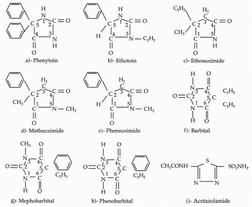
Until phenytoin was marketed as an AED in 1938, phenobarbital was well established as the agent of choice in the treatment of seizures. Merritt and Putnam (2,3), searching for AEDs devoid of sedative effects, first reported on the anticonvulsant properties of phenyl derivatives in animal studies. They recommended clinical trials of phenytoin (Dilantin; 5,5-diphenylhydantoin; Fig. 69.1A), and soon demonstrated the superiority of the agent over phenobarbital and its lack of significant hypnotic effects (4). Phenytoin has since become the world’s most commonly used agent for the treatment of patients with generalized tonic-clonic and simple and complex partial seizures. Other hydantoins were also tested, but only ethotoin is still in use today.


Chemistry and Mechanism of Action

Ethotoin (Peganone, 3-ethyl-5-phenylhydantoin; Fig. 69.1B) is similar to phenytoin, except for the deletion of one
phenyl group from position 5 and the addition of an ethyl group in position 3 of the hydantoin ring. It has a molecular mass of 204.22. Ethotoin has a broad spectrum of activity, and inhibits seizures induced by maximal electro-shock and pentylenetetrazol.


TABLE 69.1 ANTIEPILEPTIC DRUGS MARKETED IN THE UNITED STATES


Year


Nonproprietary Namea


Trade Name Introduced


1912


Phenobarbitalb


Luminal


1935


Mephobarbital


Mebraral


1938


Phenytoin


Dilantin


1946


Trimethadione


Tridione


1947


Mephenytoin


Mesantoin


1949


Paramethadione


Paradione


1951


Phenacemide


Phenurone


1952


Metharbital


Gemonil


1953


Phensuximide


Milontin


1954


Primidone


Mysoline


1957


Methsuximide


Celontin


1957


Ethotoin


Peganone


1960


Ethosuximide


Zarontin


1968


Diazepam


Valium


1974


Carbamazepine


Tegretol


1975


Clonazepam


Clonopin


1978


Valproate


Depakene


1981


Clorazepate


Tranxene


1992


Felbamate


Felbatol


1994


Gabapentin


Neurontin


1995


Lamotrigine


Lamictal


1996


Topiramate


Topamax


1997


Tiagabine


Gabitril


2000


Oxcarbazepine


Trileptal


2000


Zonisamide


Zonegran


2000


Levetiracetam


Keppra


a Withdrawn AEDs are not included.

b Boldface indicates major AEDs.


Adapted from Levy R, Mattson R, Meldrum B, et al, eds. Antiepileptic drugs, 3rd ed. New York: Raven Press. 1989:xxiv.


Absorption, Distribution, and Metabolism

Ethotoin is slowly absorbed from the gastrointestinal (GI) tract. Absorption is dose dependent; the time to peak plasma concentration increases with increasing dose. This non-linear metabolic profile may explain the poor correlation between daily dose and steady-state serum levels of ethotoin (5).

Ethotoin is metabolized in the liver by hydroxylation and deethylation of the hydantoin ring. It has a relatively short half-life of 6 to 9 hours.


Efficacy and Clinical Use

The clinical use of ethotoin has been limited by its hypnotic properties and low anticonvulsant potency (6). The lack of gingival hyperplasia and hirsutism, side effects of phenytoin therapy, may make ethotoin an attractive alternative AED; however, it is only one fourth as effective as phenytoin in inhibiting electrically induced convulsions in animals. Few clinical trials of ethotoin are cited in the literature. In one study (7), ethotoin reduced seizure frequency in most of the children (N = 17) with uncontrolled seizures treated with dosages of 19 to 49 mg/kg per day. Two hours after ingestion, serum levels ranged from 14 to 34 mg/mL (conversion for ethotoin: μmol/L = μg/mL × 4.90) (5). In a retrospective study of adults with medically refractory epilepsy, ethotoin as adjunctive therapy reduced overall seizure frequency, especially the frequency of tonic seizures (8). The efficacy of the agent, however, was reduced by one half within 10 months, suggesting rapid onset of tolerance. Ethotoin is ineffective in treating and may exacerbate absence seizures. Because of its short half-life, ethotoin is given in four divided doses of 20 to 40 mg/kg per day. Ethotoin is available in 250- and 500-mg tablets.


Interactions with Other Agents and Adverse Effects

No drug-drug interactions have been documented with ethotoin.


Figure 69.1 Chemical structures of selected minor and major antiepileptic drugs: A: Phenytoin. B: Ethotoin. C-E: Methsuximide belongs to the succinimide family (ethosuximide and phensuximide), which shares a common heterocyclic ring. D: Methsuximide. F-G: Mephobarbital is structurally similar to barbital. H: Phenobarbital. I: Acetazolamide.

Although the agent is relatively free of the common adverse effects of phenytoin, ataxia, diplopia, dizziness, insomnia, rash, and GI distress may occur during ethotoin use. Isolated cases of lymphadenopathy have been reported. Cleft lip, cleft palate, and other malformations have occurred in infants born to mothers taking ethotoin (9,10).

Ethotoin has been available for more than 3 decades, but its efficacy and safety have not been adequately established in well-controlled clinical trials, and its use in the treatment of seizures and epilepsy remains limited.


METHSUXIMIDE


Historical Background

Introduced in 1957 for the treatment of refractory absence seizures, methsuximide (Celontin2) belongs to the succinimide family (i.e., ethosuximide and phensuximide), which shares a common heterocyclic (succinimide) ring (Fig. 69.1C-E). The diverse effects of these agents in a variety of experimental and clinical seizure types are probably related to the substitution of different chemical groups in the succinimide ring. Since phensuximide is no longer available, only methsuximide is discussed in detail in this chapter.


Chemistry and Mechanism of Action

The chemical structure of methsuximide (N-2-dimethyl-2-phenyl-succinimide) is shown in Figure 69.1D. Phenyl group substitution at the 2C position counteracts experimentally induced maximal electroshock seizures, whereas alkyl group substitution at the 2C position counteracts experimentally induced pentylenetetrazol seizures. Methyl group substitution at the 5N position adds to the antipentylenetetrazol effect and the sedative activity. Alkyl substitution at the 5N and 2C positions and phenyl substitution at the 2C position provide activity against pentylenetetrazol- and maximal electroshock-induced seizure activity (11).

Methsuximide is a nonpolar chemical compound that is water soluble and slightly lipophilic. Its exact effects on excitable membranes are unknown. Because of its effectiveness against absence and partial seizures, the agent probably has more than one mechanism of action, including effects on transmitter release, calcium uptake into
presynaptic endings, and conductance of sodium, potassium, and chloride.


Absorption, Distribution, and Metabolism

Methsuximide is quickly absorbed through the GI tract, with peak plasma levels achieved in 2 to 4 hours. The agent is distributed evenly throughout the body and penetrates brain and fat tissue better than ethosuximide (12). Because of its low protein binding and poor solubility, methsuximide equilibrates with CSF (B.J. Wilder, unpublished data, 1980). It is rapidly metabolized to N-desmethyl-methsuximide or 2-methyl-2-phenyl succinimide (12, 13, 14, 15) and has a mean half-life of 1.4 hours. Trough plasma concentrations of methsuximide are reportedly undetectable in fasting specimens (16). A major active metabolite of methsuximide, N-desmethyl-methsuximide, achieves high steady-state plasma levels and exerts a major anticonvulsant effect. The mean half-life of this metabolite is 38 hours (range, 37 to 48 hours) (13), although some investigators (16) have reported half-lives of 51.6 to 80.2 hours in patients who received maximal doses of methsuximide. Another methsuximide metabolite, N-methyl-2-hydroxymethyl-2-phenylsuccinimide, was detected by means of gas chromatography and mass spectrometry of the serum of a patient with a fatal overdose of primidone and methsuximide.

Optimal clinical effect may be achieved with a nontrough N-desmethyl-methsuximide plasma concentration of 20 to 24 μg/mL (15), near the middle of the therapeutic range of 10 to 40 μg/mL reported by Strong and colleagues (14). Browne and associates (16) reported a therapeutic range of 10 to 30 μg/mL for fasting N-desmethylmethsuximide plasma concentrations. Steady-state plasma concentration is reached between 8.1 and 16.8 days from onset of maintenance methsuximide dose. The usual dosage increase of 150 or 300 mg per day can be made at biweekly intervals to avoid toxicity. Methsuximide is no longer available in 150-mg tablets; biweekly dosage increments of one tablet (300 mg) every other day may be used (16).


Efficacy and Clinical Use

Methsuximide has a wide spectrum of antiepileptic activity and is effective in patients with complex partial seizures (15, 16, 17), generalized tonic-clonic seizures, and absence seizures (18, 19, 20, 21). Wilder, Buchanan, and Uthman (15,22) found methsuximide to be an effective adjunctive agent in the management of refractory complex partial seizures. Twenty-one patients taking phenytoin, phenobarbital, primidone, or carbamazepine as monotherapy or in combination were studied. Of these patients, 71% achieved good to excellent control of complex partial seizures, and a dose reduction or discontinuation of 1 or more AED was possible in 42%. Optimal plasma levels and control of complex partial seizures were associated with daily methsuximide dosages of 9.5 to 11.0 mg/kg, with maximal seizure control observed at N-desmethyl-methsuximide plasma levels of 20 to 24 μg/mL (conversion factor for methsuximide: μmol/L = 4.92 × μg/mL). A dose-response relationship was determined after the addition of methsuximide, and seizure frequency progressively decreased as N-desmethyl-methsuximide serum levels increased.

Browne and colleagues (16) described the use of adjunctive methsuximide in 26 patients with medically refractory complex partial seizures. The maximal tolerated dose of methsuximide was maintained for 8 weeks. Of the total population, eight patients (31%) had a 50% or more reduction in seizure frequency, and four (15%) became seizure free. Eight patients withdrew from the study because of adverse events and three because of increased seizure frequency (these patients had a history of severe seizure flurries before and after initiation of methsuximide treatment). Of the eight patients who responded, five continued to have a 50% or more reduction in frequency of complex partial seizures for 3 to 34 months.

Sigler and associates (23) used methsuximide as add-on therapy in children with epilepsy refractory to first- and second-line AEDs. Forty patients (35.7%) had a 50% or more reduction in seizure frequency, and ten (8.9%) became seizure free during the short-term phase (mean, 9.1 weeks). Of the 112 patients studied, 22 (19.6%) continued to benefit from the drug (a 50% or more reduction in seizure frequency compared with baseline and absence of intolerable side effects) at long-term follow-up (mean, 3.7 years; range, 18 months to 7.1 years). In patients with good seizure control, fasting plasma levels of N-desmethyl-methsuximide were 25.3 to 44.7 mg/L (mean, 36.0 mg/L); thus, effective plasma concentrations of N-desmethyl-methsuximide in children were found to be higher than previously described. No serious or irreversible side effects were reported. Likewise, Tennison and colleagues (24) used methsuximide as add-on therapy in children with complex partial and “minor motor” seizures refractory to first- and second-line AEDs; 15 patients (60%) had a 50% or more reduction in seizure frequency, and no serious adverse events were reported.

Other reports of methsuximide as adjunctive medical therapy for complex partial seizures showed complete seizure control in 0% to 38% of patients and a 50% or more reduction in seizure frequency in 6% to 100% of patients (17,19,25, 26, 27, 28). In one study of previously untreated patients (29), seizures were controlled in 18%, and 27% had a 50% or more reduction in seizure frequency.

Results of early studies of methsuximide showed some efficacy in patients with absence seizures (19, 20, 21). In one study (20), methsuximide was used in previously untreated patients; absences were not completely controlled in any patient, and only 20% had a greater than 50% reduction in
seizure frequency. Rabe (30) reported that 10 of 16 patients became completely free of absences, and another five had seizure frequency reduced by 75%. He suggested two possible explanations for the greater effectiveness of methsuximide in his study compared with earlier work: Most of his patients had epilepsy of relatively recent onset, and he used considerably higher doses of methsuximide (1,200 to 2,100 mg per day) than did previous investigators.

Rabe also reported on the efficacy of methsuximide in four patients with juvenile myoclonic epilepsy (30). Two patients became completely free of myoclonus, and two had a reduction in frequency of at least 75%. One case report described methsuximide used with primidone to be very effective in a 17-year-old boy with drawing-induced myoclonic seizures (31). Hurst (32) described five adolescent girls with juvenile myoclonic epilepsy who became seizure free taking methsuximide; four were maintained on monotherapy.

Tolerance to the anticonvulsant effect of methsuximide develops in approximately 50% of patients treated with maximal doses, and seizure frequency returns to baseline. The low overall efficacy of methsuximide relative to that of first-line AEDs may reflect the selectively more refractory seizures in the patients studied. Failures because of toxic reactions might have occurred when the dose of methsuximide was increased too rapidly. The dose should not be increased more often than every 2 weeks in adults receiving multidrug therapy (16). Methsuximide should be considered in patients who are allergic to or whose disease is refractory to other AEDs.


Interactions with Other Agents and Adverse Effects

Methsuximide interacts with other AEDs, necessitating close monitoring of serum levels and adjustment of concurrent AED dose, especially in the face of clinical toxicity. Rambeck (33) reported that concurrent administration of methsuximide increased the mean serum concentration of phenobarbital by 37% in patients receiving this agent and by 40% in patients receiving primidone. The mean serum concentration of phenytoin increased by 78%. Patients taking phenobarbital or phenytoin had increased serum levels of N-desmethyl-methsuximide compared with patients taking methsuximide alone. These increases were attributed to competition by the drugs for a common hydroxylating enzyme system.

Conversely, the addition of methsuximide induces the metabolization of other AEDs (34,35). Methsuximide decreased the mean serum concentrations of carbamazepine (16), valproic acid (36,37), lamotrigine (35,38), and topiramate (39) when added to the treatment regimen. Methsuximide mitigated the effect of valproic acid on lamotrigine; the combination of valproic acid and lamotrigine increased the concentration of lamotrigine by 211% compared with lamotrigine monotherapy, however, if methsuximide was added, the increased concentration of lamotrigine dropped to 8% (35).

GI disturbance, lethargy, somnolence, fatigue, and headache may be experienced, but these adverse effects are usually transient and dose-related. Other adverse experiences include hiccups, irritability, ataxia, blurred vision or diplopia, inattention, dysarthria, and psychic changes (16). In some patients, headache, photophobia, and hiccups require withdrawal of methsuximide (15). Transient leukopenia and a movement disorder have been reported (40). Delayed, profound coma following methsuximide overdose has been described (41). Charcoal hemoperfusion was successful in one case of methsuximide overdose (42).


BARBITURATES


Historical Background

Approximately 2500 barbiturate compounds have been synthesized since barbituric acid was first produced in 1864. About 25 (1%) of these compounds are licensed by the U.S. Food and Drug Administration as hypnotics, anesthetics, and anticonvulsants (11). Two barbiturates are currently marketed as AEDs: mephobarbital (Mebaral3, methylphenobarbital, methylphenobarbitone); and phenobarbital (Luminal; Fig. 69.1H). Primidone (Mysoline) is a deoxybarbiturate metabolized to phenobarbital and phenylethyl-malonamide. All these compounds are derived from barbital, the first synthetic hypnotic barbiturate.

Phenobarbital was introduced as an AED in 1912 (43) and remains one of the major agents used worldwide for the treatment of generalized tonic-clonic and simple or complex partial seizures. It has a special use in patients with status epilepticus and is widely prescribed for prophylaxis of febrile seizures and alcohol- and drug-withdrawal seizures. Phenobarbital and primidone were reported to be less well tolerated than phenytoin and carbamazepine for the treatment of seizures of partial onset (44). A number of studies have reported on the behavioral and cognitive side effects of phenobarbital (45, 46, 47, 48, 49). Phenobarbital and primidone are discussed in detail in other chapters. Mephobarbital is considered here.


Chemistry and Mechanism of Action

The chemical structure of mephobarbital (5-ethyl-1-methyl-5-phenylbarbituric acid) is similar to the structure of barbital, as illustrated in Figures 69.1F and G. Mephobarbital is similar to phenobarbital except for the methyl group at the 3N position. The molecular mass of mephobarbital is 246.26. The mechanism of anticonvulsant action is
probably similar to that of phenobarbital, essentially inhibiting the spread of seizure activity and elevating seizure threshold (50). All the commercially available hypnotic barbiturates exhibit anticonvulsant activity at anesthetic doses and inhibit epileptic seizures induced by electroshock, tetanus, strychnine, or pentylenetetrazol. This anticonvulsant activity is separate from the sedative or anesthetic effects and is not diminished by the concurrent administration of agents that counteract sedation (11).


Absorption, Distribution, and Metabolism

Mephobarbital is highly soluble in lipids, with lipid to water partition ratio of 100. The agent is easily absorbed and readily crosses biologic membranes. A bioavailability of 75% was found in a pharmacokinetic study of mephobarbital in two volunteers (51). It appears to be widely distributed in the body, with higher concentrations in adipose tissue and brain. In rats, brain mephobarbital levels were 8 times those simultaneously measured in blood (52). In vitro studies suggest that 58% to 68% of mephobarbital in highly concentrated solution is bound to human serum albumin (53).

Only gold members can continue reading. Log In or Register to continue

Stay updated, free articles. Join our Telegram channel

Oct 17, 2016 | Posted by in NEUROLOGY | Comments Off on Less Commonly Used Antiepileptic Drugs

Full access? Get Clinical Tree

Get Clinical Tree app for offline access