Leukodystrophies


Leukodystrophies


Patricia L. Musolino

Florian Eichler


GENERAL CONCEPTS

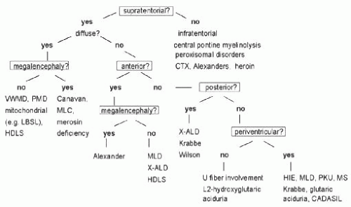
Leukodystrophies are inherited disorders that affect the white matter of the central nervous system with or without involvement of the peripheral nerve. Collectively leukodystrophies represent an important cause of progressive neurological disability in children. All disorders share glial cell or myelin sheath involvement. Brain MRI plays a large role in detection and diagnosis. Patterns of white matter involvement can be pathognomonic (Fig. 9.1). Diagnosis is important for palliative or experimental therapies that may offer benefits, as well as for reproductive counseling and family screening of currently unaffected individuals. Since the few treatments available are more effective in the early stages, timely referral to an expert is crucial.1


CLINICAL FEATURES


Age of Onset

First symptoms are usually neurological and appear, with few exceptions, in previously healthy children. Most leukodystrophies occur within a particular age group and can range in onset from early infancy to late adulthood. Within a leukodystrophy the phenotypes can vary, with more severe phenotypes usually starting in early childhood. Symptoms usually progress
slowly, with possible periods of stagnation. Phenotypes are variable depending on the severity of mutations. Starting from the age of the patient at onset of symptoms, certain types can be considered (Tables 9.1 and 9.2).


FIGURE 9.1 Algorithm for the Use of MRI Patterns in Distinguishing Disorders with White Matter Involvement (Reproduced with permission from Costello DJ, Eichler AF, Eichler FS. Leukodystrophies: classification, diagnosis, and treatment. Neurologist. 2009;15:319-328.)


TABLE 9.1 Most Common Leukodystrophies by Age of Onset of Symptoms


Leukodystrophies to Consider in Relation to Age of Patient at Onset


Patient age at onset of symptoms


Leukodystrophies (in order of probability)


Infantile (1st y of life)


Globoid cell leukodystrophy


Pelizaeus-Merzbacher disease


Canavan disease


Vanishing white matter disease


Megalencephalic leukodystrophy with cysts


Aicardi-Goutières syndrome


Hypomyelination with atrophy of the basal ganglia and cerebellum


Late infantile (1-5 y)


Metachromatic leukodystrophy


Alexander disease


Vanishing white matter disease


Megalencephalic leukodystrophy with cysts


Hypomyelination with atrophy of the basal ganglia and cerebellum


Leukoencephalopathy with brainstem and spinal cord involvement and elevated lactate


Giant axonal neuropathy type I


Juvenile


X-linked adrenoleukodystrophy


Metachromatic leukodystrophy


Vanishing white matter disease


Megalencephalic leukodystrophy with cysts Alexander disease


Leukoencephalopathy with brainstem and spinal cord involvement and elevated lactate


Adolescence, young adulthood


Metachromatic leukodystrophy


Vanishing white matter disease


Leukoencephalopathy with brainstem and spinal cord involvement and elevated lactate


Adapted from Kohlschutter A, Eichler F. Childhood leukodystrophies: a clinical perspective. Expert Rev Neurother. 2011;11:1485-1496.


Neuroimaging

MRI of the head is the most important ancillary test. Standard investigation is T1- and T2-weighted and fluid-attenuated inversion-recovery (FLAIR) images. The distribution of white matter abnormalities (see Fig. 9.1) and certain specific patterns (Table 9.3) can assist in the differentiation of leukodystrophies. A systematic assessment of lesion location, presence of hypomyelination, cystic lesions, calcifications, contrast enhancement, and abnormalities on proton MR spectroscopy (MRS) is crucial. A rough classification of leukodystrophies according to their presentation on neuroimaging is shown in Table 9.3.1,2


TABLE 9.2 Differential Diagnosis of Leukodystrophies by Age of Onset


Childhood Onset


Primary CNS inflammation (ADEM, MS)


Primary CNS infection (encephalitis)


CNS neoplasia (glial tumors, lymphoma)


Toxic leukoencephalopathy (radiation, chemotherapy, biologic therapies)


Perinatal injury (hypoxic ischemic encephalopathy, periventricular leukomalacia)


Adult Onset


Infiltrative tumors (gliomas, gliomatosis cerebri, primary CNS lymphoma)


Toxic leukoencephalopathy (radiation, chemotherapy, organic solvents, biologic therapies, drugs of abuse)


Metabolic leukoencephalopathy (anoxia, carbon monoxide, mitochondrial)


Infection (encephalitis, HIV encephalopathy, progressive multifocal leukoencephalopathy)


Trauma (diffuse axonal injury) vasculopathies (ischemic, inflammatory, CADASIL), CNS inflammation (MS, ADEM, systemic disorders with CNS involvement)


CNS indicates central nervous system; ADEM, acute demyelinating encephalomyelitis; MS, multiple sclerosis, HIV, human immunodeficiency virus; CADASIL, cerebral autosomal dominant arteriopathy with subcortical infarcts and leukoencephalopathy.


Adapted from Costello DJ, Eichler AF, Eichler FS. Leukodystrophies: classification, diagnosis, and treatment. Neurologist. 2009;15:319-328.


TABLE 9.3 Childhood Leukodystrophies According to Major Neuroimaging Patterns


Disorders with Confluent MRI Lesions


Alexander disease1


Canavan disease


Globoid cell leukodystrophy


Leukoencephalopathy with metaphyseal chondrodysplasia


Metachromatic leukodystrophy


Metachromatic leukodystrophy with multiple sulfatase deficiency


Mitochondrial disorders


X-linked adrenoleukodystrophy


Disorders with Cavitating MRI Lesions


Cystic leukoencephalopathy without megalencephaly


Glycine leukoencephalopathy


Leukoencephalopathy with calcifications and cysts


Megalencephalic leukodystrophy with cysts


Progressive cavitating leukoencephalopathy


Disorders with Hypomyelination


Fucosidosis


Folate receptor defect


Hypomyelination with atrophy of the basal ganglia and cerebellum


Hypomyelination and congenital cataract


Hypomyelination, hypodontia, hypogonadotropic hypogonadism


Hypomyelination with monocarboxylate transporter-8 deficiency


Pelizaeus-Merzbacher disease


Pelizaeus-Merzbacher-like disease


Sialic acid storage disorder


Tremor-ataxia with central hypomyelination


Disorders with Calcifications


Aicardi-Goutières syndrome


Cerebrotendinous xanthomatosis


Leukoencephalopathy with calcifications and cysts


1 Infantile type.


Adapted from Kohlschutter A, Eichler F. Childhood leukodystrophies: a clinical perspective. Expert Rev Neurother. 2011; 11:1485-1496.


Physical and Neurological Exams

Most patients with leukodystrophies do not have any physical abnormalities. Some have a large head (Alexander disease, Canavan disease, megalencephalic leukodystrophy with cysts, and vanishing white matter disease). Very rarely, they can have dysmorphic features and skeletal abnormalities that resemble those seen in mucopolysaccharidoses (fucosidosis and metachromatic leukodystrophy [MLD] with multiple sulfatase deficiency). Dental abnormalities are seen in some forms of hypomyelination, such as hypomyelination, hypodontia, and hypogonadotropic hypogonadism (4H syndrome).1


MOST COMMON LEUKODYSTROPHIES (TABLE 9.4)


X-linked Adrenoleukodystrophy (ALD)

Defect: Mutations in the ABCD1 gene lead to defects in peroxisomal betaoxidation & very long chain fatty acid (VLCFA) accumulation. Locus Xq28.

Presentation: Cerebral ALD (only seen in males): Onset 4 to 8 y, 85% present initially with neurologic symptoms. Often begins with personality changes (resembles ADHD) and academic performance decline, followed by ataxia, spasticity, loss of vision and hearing. Dementia and seizures can occur later in the course. It often leads to total disability within 2 y. Adrenomyeloneuropathy (AMN) or adult form (males and some females): Onset after 20 y with spastic paraplegia, sensory peripheral neuropathy, sphincter disturbances, and impotence. Other si/sx: Adrenal insufficiency (mean onset 7.5 y, may be isolated). Prognosis: In cerebral ALD, there is progression to vegetative state and death by 3 y after onset (slower progression in adult cerebral ALD). FHx: X-linked, only males affected in cerebral form, up to 20% of female carriers will develop AMN.

Jun 20, 2016 | Posted by in NEUROLOGY | Comments Off on Leukodystrophies

Full access? Get Clinical Tree

Get Clinical Tree app for offline access