Magnetic Resonance Imaging Techniques in the Evaluation for Epilepsy Surgery


Magnetic Resonance Imaging Techniques in the Evaluation for Epilepsy Surgery


Susanne Knake

P. Ellen Grant


Approximately 60% of all patients with epilepsy (0.4% of the population in industrialized countries) suffer from focal epilepsy syndromes. About 15% of these patients have medically intractable seizures and might be candidates for surgical epilepsy treatment (1,2). The goal of surgical treatment is seizure control by complete disconnection of the epileptogenic zone, which is defined as the area of tissue indispensable for the generation of clinical seizures under preservation of the eloquent cortex (2). Magnetic resonance imaging (MRI) enables the detection of anatomic lesions that are candidates for the epileptogenic zone (3). A focal anatomic abnormality involving cortex (either neocortex or hippocampus) that is identified in a region consistent with clinical semiology or electroencephalographic (EEG) findings is the probable cause of the epileptic seizures (4). In temporal lobe epilepsy, the probability of a seizure-free outcome is 82% with concordant lesions and 56% with an unremarkable MRI (5). Respective figures in frontal lobe epilepsy are 72% and 41% (6). MRI detects surgically relevant lesions in up to 80% of patients who undergo temporal lobectomy (6, 7, 8, 9) and in about 60% of those undergoing frontal lobe surgeries (6). MRI also detects cerebral malformations associated with epilepsy syndromes, thereby helping to guide medical therapy. When coregistered, MRI can illustrate the topographic relationship between lesions and eloquent cortex defined by functional MRI, EEG, single-photon-emission computed tomography (SPECT), positron emission tomography (PET), or magnetoencephalography (MEG).

This chapter reviews the criteria for use of an MRI and the type of MRI that should be used, outlines the imaging sequences to evaluate mesial temporal and neocortical epilepsies, and describes the imaging findings in associated conditions.


WHO SHOULD HAVE AN MRI?

The International League Against Epilepsy recommends one nonemergent MRI for all patients except those with idiopathic generalized epilepsies (10,11). MRI is often normal in IGE (12,13), but most of these studies did not include high-resolution techniques. In addition, the differences between focal epilepsy and IGE may not be distinct, and some seizures that appear to be generalized by clinical and EEG criteria are actually rapidly spreading partial seizures (14). Because MRI might help to exclude an unusual but potentially treatable cause, one state-of-the-art examination may be reasonable in every epilepsy patient.


WHAT TYPE OF MRI SHOULD BE ORDERED?

MRI should be performed on a state-of-the-art 1.5- or 3-T scanner. The poor image quality of low-field or open systems could miss subtle abnormalities despite interpretation by experienced neuroradiologists. Even if performed
on a 1.5-T system, older MRI techniques may not include all of the modern sequences. Phased-array imaging at 1.5 T or 3 T, can increase lesion detection and should be considered for patients with focal epilepsies, particularly medically refractory seizures, in whom 1.5-T studies with a standard head coil yield normal or discordant results (15,16). The imaging sequences performed will depend on the duration and type of epilepsy as well as the age of the patient because the differential diagnosis differs in each of these categories (Table 74.1). The protocol for new-onset seizures in adults typically includes postcontrast studies; in children, whose seizures are less often secondary to neoplasms, a high-resolution protocol with contrast is used only on identification of a lesion. High-resolution T1-weighted and T2-weighted images through the hippocampi are recommended for mesial temporal lobe epilepsy. The same type of weighted images are appropriate for focal neocortical epilepsies but through the lobe of interest if no lesion is obvious and including in adults a gradient-echo T2 sequence. Therefore, when MRI studies are requested, new-onset seizures should be distinguished from epilepsy syndromes and the type of epilepsy should be described. Imaging protocols are discussed below.


TABLE 74.1 3T PROTOCOLa


Sequence


Direction


Slices (no.)


TR (ms)


TE (ms)


Matrix (mm)


Slice Thickness (mm)


Gap (%)


Min (˜)


iPAT


MPRAGE


Sagittal (coronal and axial reformats)


160


2000


3.71


256 × 256


1


0


4:40


2


High-resolution T2 TSE


Coronal or axial


27


6000


96


512 × 384


3


0


6:08


0


FLAIR


Coronal or axial


28


10,000 TI = 2500


70 10 degrees


384 × 288


5


20


3:32


0


Gradient echo T2


Coronal or axial


24


359


12


256 × 252


5


0


3:21


2


aIn addition to routine brain sequences, for patients with focal seizures. For mesial temporal sclerosis, the high-resolution T2 and FLAIR sequences should be angled perpendicular to the axis of the hippocampi. For neocortical epilepsy, the optimal imaging plane for the lobe of interest is chosen. Abbreviations: FLAIR, fluid-attenuated inversion recovery; MPRAGE, magnetization-prepared rapid gradient echo; TE, echo time; TI, inversion time; T2 TSE, T2-turbo-spin-echo; TR, repetition time; iPAT, integrated parallel acquisition technique.


MESIAL TEMPORAL LOBE EPILEPSY

A tailored MRI protocol is important for diagnosing mesial temporal sclerosis (MTS) and predicting surgical outcome. Assessment by an experienced observer is also crucial (17) as MTS especially is often missed by the inexperienced reader (18). Radiographically proven unilateral hippocampal sclerosis is closely correlated with seizure-free outcome in up to 80% of patients after temporal lobe resections (5,19, 20, 21, 22). MRI also detects dual pathology, which occurs in about 15% of patients with hippocampal sclerosis (23), involves a less favorable outcome, and usually requires resection of both abnormalities for control (24,25). Dual pathology is common in patients with developmental disorders of the temporal lobe and may be associated with bilateral hippocampal abnormalities (26,27).


Protocol

High-resolution images through the hippocampi can detect the primary findings that establish a diagnosis of MTS as well as secondary abnormalities (Table 74.1). Images through the rest of the brain allow detection of dual pathology. Although imaging sequences may vary slightly from center to center, essential components are thin-section high-matrix T2 fast spin echo (FSE), thin-section fluid-attenuated inversion recovery (FLAIR) images angled perpendicular to the long axis of the hippocampus, and volumetric T1-weighted images with a partition size not exceeding 2 mm through the entire brain; a gradientecho T2 sequence is a high priority in adults. Symmetric alignment of the head improves the accuracy of visual assessments of asymmetric hippocampal volume. Some centers obtain short tau inversion recovery (STIR) sequences because of the excellent gray-white contrast and ability to assess the hippocampal signal. Our volumetric T1-weighted sequence provides higher resolution of the gray-white junction, and T2 FSE and FLAIR images evaluate the hippocampal signal. Spin-echo T2-weighted sequences are probably more sensitive to subtle increases in T2 signal but the long imaging times often necessitate cardiac gating to obtain adequate images.


Qualitative Assessment

The assessment usually begins with a close inspection of the hippocampi on oblique T2 FSE images, preferably with
magnification. Increased T2 signal, which reportedly has a sensitivity of 93% and a specificity of 74% (28), can be detected in specific sectors, along with blurring of the internal architecture (Fig. 74.1). In more severe cases, decreased undulations in the hippocampal head can often be identified (29). T2-signal increases in the collateral white matter and blurring of the gray-white junction are also visible on this sequence. Table 74.2 summarizes the radiographic findings of MTS (5,9,29,30, 31, 32, 33, 34, 35, 36, 37, 38, 39, 40, 41, 42, 43, 44).


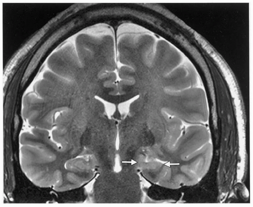
Figure 74.1 Coronal T2 FSE obtained with an 8-channel phasedarray coil at 3T. The left hippocampus is smaller than the right, T2 signal is increased, and the internal architecture is blurred (arrows). These are primary imaging features of mesial temporal sclerosis.

Close inspection of the FLAIR images confirms T2 signal asymmetry. On FLAIR images, the hippocampus normally is brighter than neocortex, and the choroid plexus just superior to the hippocampi is bright. A comparison with T2 FSE images can avoid misdiagnosing bilateral disease or mistaking choroid plexus for hippocampal hyperintensity. Scrutiny of the coronal volumetric T1 images allows comparison of hippocampal and temporal lobe volumes. Secondary MTS findings such as volume loss in the circuit of Papez are best detected on this sequence, as are temporal lobe polymicrogyria and heterotopia. A gradient-echo T2 sequence may also detect dual pathology such as subtle cavernous venous, or cryptic venous, malformations or old hemorrhagic shear injury that would otherwise be missed. A normal or small T2-bright hippocampus makes the diagnosis in the appropriate clinical setting. In asymptomatic individuals, an increased T2 signal in the hippocampus (45,46) and a small hippocampus without signal abnormality (45) have uncertain significance. Increased hippocampal volume associated with increased T2 signal may suggest acute swelling because of recent severe seizure activity (47,48), infiltrating glioma, or cortical dysplasia. Although such acute swelling can progress to MTS, the differential diagnosis of an enlarged hippocampus does not include MTS (48,49). Normal variants not associated with seizures are hippocampal sulcal remnants and choroidal fissure cysts (29,50,51); both are equal to cerebrospinal fluid in signal
intensity on all sequences. Occurring in about 10% of the population, hippocampal sulcal remnants are 1- to 2-mm structures between the dentate gyrus and the cornu ammonis that represent a failure of normal embryogenic involution of the hippocampal sulcus. Choroidal fissure cysts, located just above the hippocampus, become more frequent with age (29,51).


TABLE 74.2 PRIMARY AND SECONDARY RADIOGRAPHIC FINDINGS OF MESIAL TEMPORAL SCLEROSIS


Finding


Primary


Increased hippocampal signal intensity (5,9,30)


Hippocampal atrophy (31, 32, 33, 34)


Loss of the internal architecture (33,35, 36, 37)


Secondary (temporal lobe)


Thinning of collateral white matter in adjacent parahippocampal gyrus (38)


Loss of hippocampal head digitizations (29)


Diminished demarcation between gray and white matter in temporal lobe neocortex (38, 39, 40)


Temporal lobe atrophy and enlarged temporal horn (41)


Secondary (extratemporal)


Atrophy of the mamillary body (42)


Atrophy of fornix/amygdale (43)


Thalamic and caudate atrophy (44)


Magnetic Resonance Spectroscopy

MRS noninvasively measures the integrity and function of neuronal tissue; 31P-MRS evaluates cerebral energetics and pH. Studies at 1.5 T and 4.1 T have shown reductions of 50% in the ratio of phosphocreatine/inorganic phosphate (PCr/Pi) in the affected lobe of patients with temporal lobe epilepsy and of 24% in the contralateral temporal lobe (52, 53, 54), thought to indicate persistent impairment of energy metabolism. Despite these intriguing changes, a low signal-to-noise ratio, poor spatial resolution, and widespread unavailability for routine clinical studies limit the clinical application of 31P-MRS. 1H-MRS, in contrast, can be performed on most clinical 1.5-T scanners with spatial resolutions between approximately 1 and 8 cc, depending on whether multivoxel or single-voxel techniques are used. 1H-MRS provides biochemical information about neuronal function (N-acetyl aspartate [NAA]), membrane turnover (choline), and total energy stores (creatine [Cr]) as well as the presence of cerebral lactate. Its sensitivity and specificity in temporal lobe epilepsy ranges from 60% (55) to 97% (56) and is greater with higher-spatial-resolution techniques (57). High-quality routine MRI of the hippocampus and 1H-MRS for localization have not been directly compared but are probably complementary. Because absolute quantification of NAA, choline, and Cr is difficult, the sum of creatine and phosphocreatinine is assumed to be constant in adults and ratios of NAA and choline to Cr are usually calculated. Ipsilateral decreases in NAA/Cr occur in about 90% of MTS cases, and the side of functional abnormality corresponds highly with the side of seizure onset on EEG (58). The loss of NAA and the decreased NAA/Cr ratio are associated with dysfunctional mitochondrial metabolism and neuronal loss (38,39,59, 60, 61) but reflect neuronal as well as glial function (62). Combined with MRI findings, MRS improves predictions of surgical outcomes (63) and may help with lateralization in MRI-negative patients with temporal lobe epilepsy (64). With voxels typically including at most only part of the hippocampus, these MRS findings probably characterize the extrahippocampal, temporal lobe metabolic abnormalities in patients with mesial temporal lobe epilepsy. In fact, although NAA/Cr levels and ipsilateral hippocampal volumes can be statistically related, they share only a small percentage of variance and may be related but distinct (65,66). Temporal lobe MRS findings may also indicate a more widespread underlying pathologic condition (67). NAA/Cr decreases in the contralateral hemisphere may predict a poor surgical outcome (68). Lactate levels are normally absent in the brain or present in very low levels. The lactate resonance consists of two distinct peaks at 1.3 ppm and indicates a disturbance of the cellular oxidative mechanism. Lactate has been identified in the region of seizure activity during status epilepticus and epilepsia partialis continua and in the hippocampus within 24 hours after temporal lobe seizures (69, 70, 71, 72). Lactate may also alter the excitability of local neurons (73).

Because acute and transient changes have also been reported in MRS (71,74), results should be interpreted in conjunction with recent seizure activity. The 1H-MRS signal also may be influenced by antiepileptic medication and even by metabolic interventions such as a ketogenic diet (57,75,76).


Quantitative Assessment

Hippocampal volumetry and T2-relaxometry show increased sensitivity over visual analysis in the detection of hippocampal sclerosis ranging from about 80%-90% to 90%-95% (29,38,39,60,77, 78, 79). However, T2-relaxometry requires an additional imaging sequence that lengthens an already protracted protocol, and both techniques require manual segmentation of the hippocampus and post-processing to calculate T2 values and volumes. Although the added sensitivity has not yet prompted their incorporation into clinical protocols for mesial temporal lobe epilepsy, these techniques have enhanced our understanding of MTS. Hippocampal volumetry has disclosed a spectrum of volume loss in MTS (80,81), and total loss of volume has been correlated with reduced neuronal density (82, 83, 84). Longitudinal volumetric MRI studies have shown progressive hippocampal volume loss in patients with intractable mesial temporal lobe seizures, whereas patients with new-onset well-controlled temporal lobe epilepsy exhibited no change over the same period (85). No volume loss had been identified in extratemporal epilepsy, emphasizing the specificity of volumetric studies. Automated methods for calculating hippocampal volumes are being developed (86,87) and, if validated, could be used routinely.

T2-relaxometry measures the decay in signal intensity at different echo times in a series of T2-weighted images acquired at the same slice. Techniques range from the use of a 16-echo sequence (88,89) or two echoes (90, 91, 92) and time-efficient sequences (79,91); all reliably measured the T2 signal in the hippocampus and the amygdala (93). Boundaries with cerebrospinal fluid must be carefully avoided in this operator-dependent technique. During measurement of the T2-relaxation time along the long axis of the hippocampus in healthy individuals, a study described a characteristic profile that is disturbed in patients with hippocampal sclerosis (94). The hippocampal relaxation time correlates well with the glial and neuronal ratio, particularly in the CA1 subregion (80,84).


New MRI Techniques for Temporal Lobe Epilepsies


Diffusion-Weighted Imaging

Apparent diffusion coefficients (ADC) are elevated interictally in the involved hippocampus and to a lesser extent the uninvolved hippocampus (63,95). Injection of flumazenil significantly decreases the ADC in the seizure-onset zone, as defined by EEG and structural MRI (96). If imaging was performed within 15 minutes of a seizure lasting at least 60 seconds, the most pronounced ADC decreases (up to 25% to 31%) were observed in the epileptogenic zone but were only transient (97). In prolonged status, more persistent decreases may correlate with hippocampal injury and the development of MTS (47,98).


Hippocampal Shape Analysis

Hippocampal shape analysis may suggest a typical three-dimensional pattern of volume loss in patients with mesial temporal epilepsy (99).


Arterial Spin Labeling Perfusion MRI

Arterial spin labeling (ASL) uses electromagnetic fields to label the nuclear spins of hydrogen in the water of inflowing blood, making blood itself an endogenous tracer (100). In patients with temporal lobe epilepsy, ASL can show interictal asymmetries in mesial temporal lobe cerebral blood flow (100,101); its role in neocortical epilepsy is unclear. Significant improvements in signal-to-noise ratio (SNR) at higher field strengths will optimize this technique (102).


FOCAL NEOCORTICAL EPILEPSIES

Less common than mesial temporal epilepsy in adults, focal neocortical epilepsy involves more variable and subtle abnormalities that may be located anywhere in the neocortex. For this reason, routine MRI is often interpreted as normal in neocortical epilepsy.

MRI plays a central role. Although a pathologic substrate portends a poor long-term response to medical treatment (103), a lesion corresponding to the ictal-onset zone may improve surgical prognosis. For example, 72% of patients with identifiable lesions have good surgical outcomes compared with 41% of patients with no lesions (6). Surgical outcomes improve if the entire lesion is resected, making precise anatomic delineation important (2). Evaluation of the images by an experienced reader is also critical, particularly in the absence of an obvious lesion (18). Surgical success is much lower than in mesial temporal epilepsy, often because brain MRI may show no lesion or one too extensive for complete resection (104). Among neocortical lobe epilepsies, temporal and frontal lobe syndromes are most common, followed by parietal and occipital lobe epilepsy (105,106). Frontal lobe epilepsy is less frequently treated surgically than temporal lobe epilepsy—between 5.5% and 18% in large series (107,108).

Given the large number of MRI-negative studies in neocortical epilepsy, the priority becomes thin-section imaging, with contrast reserved for new-onset epilepsy. Lengthy protocols give rise to movement artifact, degrading image quality; therefore, the study must be tailored. The structural anomalies associated with chronic partial neocortical epilepsy (107) can be grouped into mass lesions, encephalomalacia, and malformations of cortical development (MCD).

This section describes our neocortical imaging protocol and characterizes the most common lesions in the three categories. Discussions of normal imaging, the relationship between MRI-detected lesions and ictal onset, and future perspectives follow.


Protocol

Essential components are 3-mm or less high-matrix T2 FSE images obtained through the lobe of highest suspicion, coronal volumetric T1-weighted images no larger than 2 mm through the entire brain, and a gradient-echo T2 sequence (Table 74.1). Contrast should be used in all adults and in children with obvious lesions, but if time is an issue, the high-resolution sequences take precedence. A patient can always return for a contrast study if a small lesion is identified; however, if omitting the thin sections misses a small nonenhancing lesion, the disease may be labeled nonlesional. Positioning is important; symmetric alignment of the head enhances the visual assessments of gyral asymmetries. STIR sequences may be obtained, but our center uses a volumetric T1-weighted sequence to evaluate the gray-white junction. Spin-echo T2-weighted sequences can detect a subtle increased signal in the cortex or white matter associated with cortical dysplasias, but time constraints preclude their routine use.

Oct 17, 2016 | Posted by in NEUROLOGY | Comments Off on Magnetic Resonance Imaging Techniques in the Evaluation for Epilepsy Surgery

Full access? Get Clinical Tree

Get Clinical Tree app for offline access