Malformations of Cortical Development and Epilepsy


Malformations of Cortical Development and Epilepsy


William B. Dobyns

Ruben Kuzniecky


Development of the human brain is a complex process that begins with appearance of the notochord and continues long after birth (1,2). Any disruption of this process, whether by genetic or environmental factors, may result in malformations, including those of the cerebral cortex (3). Until the advent of high-resolution neuroimaging, these disorders were the domain of the pathologist. Magnetic resonance imaging (MRI) has allowed malformations of cortical development to be identified in life, leading to improved diagnosis, knowledge of their clinical consequences, and rapid progress in understanding their pathogenesis. This chapter reviews common cortical malformations associated with epilepsy.


EPIDEMIOLOGY

The incidence of malformations of cortical development has not been adequately studied, but 15% of adults and 25% of children referred to epilepsy centers for intractable partial-onset seizures have focal cortical dysplasia (4,5). The most severe cortical malformations are less common, but current estimates are probably too low, as most data predated MRI. A case ascertainment study of lissencephaly in the Netherlands showed a prevalence of 11.7 per million births (6). Data from metropolitan Atlanta from 1994 through 1999 show rates of 0.04 per 1000 for lissencephaly and 0.01 per 1000 for polymicrogyria (Birth Defects and Genetic Diseases Branch personnel, Metropolitan Atlanta Congenital Defects Program, Centers for Disease Control and Prevention, Atlanta, GA, personal communication, 2002). No data were available for other malformations of cortical development.


CLASSIFICATION

Many different malformations of cortical development, including misplaced cortical elements such as heterotopia, have been recognized. Periventricular nodular heterotopia, lissencephaly, and focal cortical dysplasia are widely known; however, other major types exist, and many new syndromes have been described.

The nomenclature and classification of these disorders continue to evolve. Although often called neuronal migration disorders, many of these malformations involve abnormal cell formation in the ventricular zone before migration or abnormal cortical organization after migration has been largely completed. A classification system (Table 3.1) was devised and subsequently modified based on fundamental embryologic and genetic principles supplemented by gross pathologic, histologic, and neuroimaging criteria (7,8). The classification system will likely change further as the diagnoses and pathogenetic mechanisms of these malformations of cortical development are refined.


MALFORMATIONS OF CORTICAL DEVELOPMENT

Accurate diagnosis relies on recognition of the malformation on brain imaging studies; assessment of the prognosis and genetic counseling both depend on the specific diagnosis. In the following sections the major groups are reviewed, including some specific syndromes (Table 3.2) with their causative genes (2,4,9, 10, 11, 12, 13, 14, 15, 16, 17, 18, 19, 20, 21, 22, 23, 24, 25, 26, 27, 28, 29, 30, 31, 32, 33, 34, 35, 36, 37, 38, 39, 40, 41, 42, 43, 44, 45, 46, 47, 48, 49, 50, 51, 52, 53, 54, 55, 56, 57, 58, 59, 60, 61, 62, 63, 64, 65, 66, 67, 68, 69, 70, 71, 72, 73, 74, 75, 76, 77, 78, 79, 80, 81, 82, 83, 84, 85, 86, 87, 88, 89, 90, 91, 92, 93, 94, 95, 96, 97, 98, 99, 100, 101, 102, 103, 104, 105, 106, 107, 108, 109, 110, 111, 112, 113, 114, 115, 116, 117, 118, 119).


TABLE 3.1 CLASSIFICATION OF BRAIN MALFORMATIONSa


  1. Malformations caused by abnormal neuronal and glial proliferation or apoptosis


    1. Decreased proliferation or increased apoptosis—microcephalies


      1. Microcephaly with normal to thin cortex


      2. Microlissencephaly (extreme microcephaly with thick cortex)


      3. Microcephaly with polymicrogyria or other cortical dysplasia


    2. Increased proliferation or decreased apoptosis (normal cell types)—megalencephalies


    3. Abnormal proliferation (abnormal cell types)


      1. Nonneoplastic


        1. Cortical hamartomas of tuberous sclerosis


        2. Cortical dysplasia with balloon cells


        3. Hemimegalencephaly


      2. Neoplastic (associated with disordered cortex)


        1. Dysembryoplastic neuroepithelial tumor


        2. Ganglioglioma


        3. Gangliocytoma


  2. Malformations caused by abnormal neuronal migration


    1. Lissencephaly and subcortical band heterotopia spectrum


    2. Cobblestone brain malformation with congenital muscular dystrophy


    3. Heterotopia


      1. Subependymal (periventricular) heterotopia


      2. Subcortical heterotopia (other than band heterotopia)


      3. Marginal glioneuronal heterotopia


  3. Malformations caused by abnormal cortical organization (including late neuronal migration)


    1. Polymicrogyria and schizencephaly


      1. Bilateral polymicrogyria syndromes


      2. Schizencephaly (polymicrogyria with clefts)


      3. Polymicrogyria with other brain malformations or abnormalities


      4. Polymicrogyria or schizencephaly as part of multiple congenital anomaly/mental retardation syndromes


    2. Cortical (architectural) dysplasia without balloon cells


    3. Microdysgenesis


  4. Malformations of cortical development, not otherwise classified


    1. Malformations secondary to inborn errors of metabolism


      1. Mitochondrial and pyruvate metabolic disorders


      2. Peroxisomal disorders


    2. Other unclassified malformations


      1. Sublobar dysplasia


      2. Others


aEach main category is expanded in additional tables in Reference 8.


MALFORMATIONS OF NEURONAL AND GLIAL PROLIFERATION WITH ABNORMAL CELL TYPES

Malformations in this group are characterized by abnormal neurons and, often, glia. Increased cell numbers further support abnormal proliferation early in development. All of these are localized malformations; in some, abnormal cell types have been classified as neoplastic, although the malignant potential is low. The most common of these is tuberous sclerosis, which is reviewed in chapter 36.


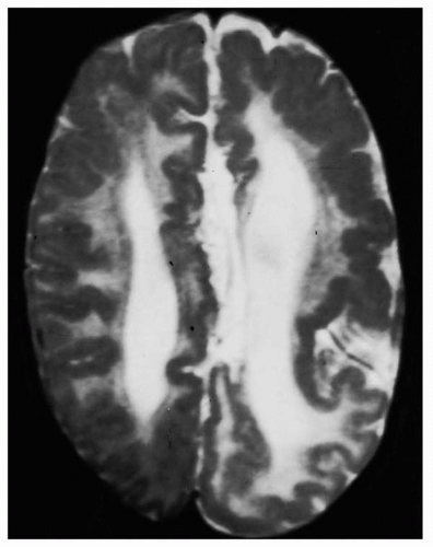
Figure 3.1 Hemimegalencephaly. Axial T2-weighted MRI shows hemimegalencephaly associated with an irregular surface and apparent pachygyria (probably fused polymicrogyria) of the entire left hemisphere and part of the right frontal region. White matter signal is strikingly increased on both sides, although more severe on the left. This child was reported previously. (From Dodge NN, Dobyns WB. Agenesis of the corpus callosum and Dandy-Walker malformation associated with hemimegalencephaly in the sebaceous nevus syndrome. Am J Med Genet 1995;56:147-150, with permission.)


Hemimegalencephaly

Patients with hemimegalencephaly have mental retardation and, typically, intractable epilepsy. The overgrowth may involve part of one hemisphere, an entire hemisphere, or an entire hemisphere and part of the other side. Pathologic changes include those seen in cortical dysplasias occurring elsewhere in the brain, white matter abnormalities, and abnormal cell types. Although most patients have no other congenital anomalies, hemimegalencephaly has been observed in several sporadic neurocutaneous syndromes and rarely in tuberous sclerosis, an autosomal dominant disorder (Table 3.2). Recurrence in families has never been reported.

The clinical presentation almost always includes seizures that usually start within the first 6 months of life and arise from the enlarged and dysplastic hemisphere (120). The seizures are typically partial, with secondary generalization, and often intractable to medical therapy. Infantile spasms
and drop attacks may present in early childhood. Unilateral neurologic signs, such as hemiparesis and hemianopia, are common. Minimal neurologic dysfunction with normal cognitive abilities has been reported in some patients, when less than a full hemisphere is involved.


Table 3.2 KNOWN CORTICAL MALFORMATION SYNDROMES, GENES, AND LOCI


Malformations and Syndromes


Genes, Loci


Hemimegalencephaly (HMEG)


HMEG, isolated


None known


Epidermal nevus syndrome (9,10)


None known


Hypomelanosis of Ito (11)


None known


Klippel-Trenaunay syndrome


None known


Neuromelanosis (12)


None known


Proteus syndrome (13)


None known


Tuberous sclerosis (14, 15, 16, 17)


TSC1, TSC2


Focal cortical dysplasia (FCD) with balloon cells (18, 19, 20)


None known


Focal transmantle dysplasia


None known


Megalencephaly (MEG)


MEG, isolated (21)


None known


Macrocephaly-CMTC syndrome (22,23)


None known


MEG with mega-corpus callosum (24)


None known


MEG-PMG-polydactyly-hydrocephalus (unpublished data) (200)


None known


Microcephaly (MIC), moderate phenotype


MIC group 1 (25, 26, 27, 28, 29, 30, 31, 32, 33, 34)


ASPM, MCPH1, 9q34, 15q, 19q13


MIC, severe phenotype


Amish lethal MIC (35,36)


SLC25A19


MIC with heterotopia (37,38)


ARFGEF2


MIC group 2, other types (39, 40, 41, 42)


None known


Seckel syndrome (43)


ATR


Microlissencephaly (MLIS)


MLIS group a, a=p (44, 45, 46)


None known


Barth MLIS syndrome (group b), a=p (45,47,48)


None known


Periventricular nodular heterotopia (PNH)


X-linked PNH (females) (49, 50, 51)


FLN1


PNH with agenesis corpus callosum (males) (52,53)


None known


PNH with Ehlers-Danlos syndrome (females) (54,55)


None known


PNH with frontonasal malformation (males) (56,57)


None known


PNH with mental retardation (males) (58,59)


None known


PNH with nephrosis (males) (60)


None known


PNH with short gut syndrome (males) (2,61)


Xq28 (?)


PNH with frontoperisylvian PMG (unpublished data)


None known


PNH with posterior-inferior PMG (unpublished data)


None known


PNH with mental retardation and duplication 5p (62)


5p15.1, 5p15.33


Heterotopia, other types


Periventricular laminar heterotopia (unpublished data)


None known


Subcortical nodular heterotopia (63)


None known


Classic lissencephaly


Baraitser-Winter syndrome, a>p (64, 65, 66, 67)


None known


Miller-Dieker syndrome a=p (44,68,69)


LIS1, 14-3-3ε


Isolated LIS sequence, a=p, a>p (70,71)


DCX


Isolated LIS sequence, a=p, p>a (70, 71, 72, 73, 74, 75)


LIS1


Subcortical band heterotopia a=p, a>p (76, 77, 78, 79, 80)


DCX


Subcortical band heterotopia, p>a (66,68,70,71,79)


LIS1


LIS with cerebellar hypoplasia (LCH)


LCH group a, a=p, a>p, p>a (81)


LIS1, DCX


LCH group b, a>p (81, 82, 83)


RELN


LCH group d, a=p (81)


None known


LIS with agenesis of the corpus callosum (ACC)


X-linked LIS with abnormal genitalia (XLAG), p>a (84, 85, 86)


ARX


LIS with ACC, other types, a=p (87, unpublished data)


None known


Cobblestone malformations


Fukuyama congenital muscular dystrophy (88, 89, 90, 91)


FCMD


Muscle-eye-brain disease, a>p (92, 93, 94, 95, 96)


POMGnT1, FKRP, LARGE


Walker-Warburg syndrome, a=p (97, 98, 99)


POMT1, FCMD


Excessive neurons in white matter (100,101)


None known


Polymicrogyria (PMG), regional


Frontal PMG (102)


None known


Frontoparietal PMG (103, 104, 105)


GPR56


Parasagittal PMG (unpublished data)


None known


Perisylvian PMG (106, 107, 108, 109, 110, 111, 112, 113)


1p36, 1q44, 22q11, Xq28


Medial parietooccipital PMG (114)


None known


Generalized PMG (115)


None known


Polymicrogyria (PMG), others


PMG with abnormal white matter (unpublished data)


None known


Schizencephaly (116)


None known


Septooptic dysplasia-schizencephaly (117, 118, 119)


None known


FCD and related


FCD without balloon cells (20)


None known


Microdysgenesis (4)


None known


a=p, Anterior equals posterior gradient; a>p, anterior more severe than posterior gradient; p>a, posterior more severe than anterior gradient; CMTC, cutis marmorata telangiectatica congenita.

Only gold members can continue reading. Log In or Register to continue

Stay updated, free articles. Join our Telegram channel

Oct 17, 2016 | Posted by in NEUROLOGY | Comments Off on Malformations of Cortical Development and Epilepsy

Full access? Get Clinical Tree

Get Clinical Tree app for offline access