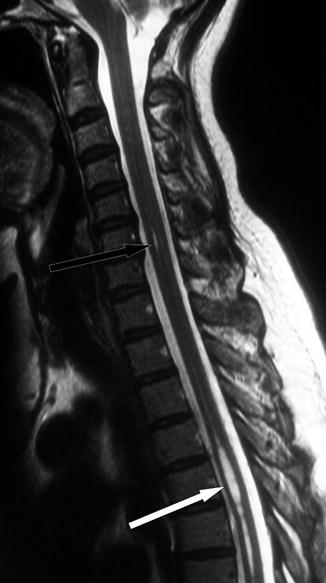
Fig. 19.1
MR image of a “cleft” or “spindle ”. This T2-weighted midline sagittal MRI of the cervical spine shows a typical cleft (long arrow) behind the body of C7. The craniovertebral junction is normal (short arrow)

Fig. 19.2
MR image of a focal dilatation of the central canal . This T2-weighted midline sagittal MRI of the cervical and upper thoracic spine shows a short, persisting segment of the central canal behind C6 (dark arrow) but a much more prominent dilatation of the central canal in the upper thoracic cord, extending over several segments from T5 downwards (white arrow). The craniovertebral junction is normal
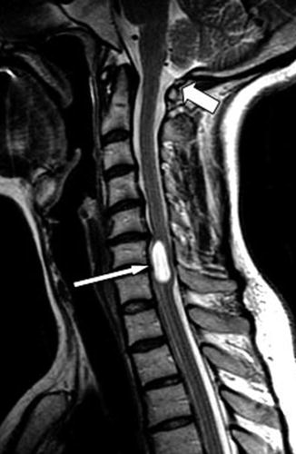
Another type of intramedullary cavity, which also extends over just a few segments of the cord, has a more rounded appearance (Fig. 19.3). Some authorities regard these lesions as being glioependymal cysts and distinct from syringomyelia (Saito et al. 2005). Exploring such a lesion will simply confirm that the appearance of the contained fluid is that of normal CSF, within otherwise normal cord tissue. Biopsy of the lining will reveal the presence of normal ependymal cells.


Fig. 19.3
MR image of a probable glioependymal cyst . This T2-weighted midline sagittal MRI of the cervical spine reveals a short, “plump”, CSF-filled cavity within the spinal cord (long arrow). The craniovertebral junction is normal (short arrow). The disc protrusion at the upper aspect of this lesion is unlikely to be related, and this appearance most likely represents that of a glioependymal cyst, although contrasted images are needed to exclude an underlying neoplasm
Whether or not these various lesions are separate conditions or part of the overall spectrum of syringomyelia, they all pose the same question as to just why they exist in the first place and why they fill with cerebrospinal fluid – or at least a fluid with similar characteristics to CSF.
From the medicolegal perspective, the question arises as to whether clefts, spindles or focal dilatations of the central canal, when detected, should be regarded as incidental findings or whether they could be generating the patient’s symptoms and whether, indeed, they could have arisen as a result of an accident in question. Many experts will declare that spinal cord clefts or spindles are not caused by or related to trauma, but others may find it difficult to disregard the existence of a relatively rare lesion, in the presence of neurological symptoms, whose onset bears a close temporal relationship to the accident in question. On the other hand, the type of symptoms as may be encountered in such cases is often equally consistent with a radiculopathy in the arms. This provides an alternative explanation that the much more common degenerative disc disease, which was previously silent, has been rendered symptomatic by the injury in question.
19.3 Medical Negligence
The purpose of this section is not to advise patents and lawyers when to take legal action over surgery, which has proven unsuccessful or led to complications. Nor is it to tell surgeons how to avoid becoming involved in litigation. Instead, we wish to highlight some of the commonly recognised complications of syringomyelia surgery, so that patients can be more fully informed, by the surgeon, before they agree to undergo an operation. It is also hoped that the surgeon might be better prepared to avoid some complications and to deal more effectively with those that do arise. The section considers, for the most part, unwanted outcomes following surgery, rather than errors in diagnosis.
In the United Kingdom, medical practitioners are regulated by the General Medical Council, and their guidance booklet “Good Medical Practice” underpins all professional activity (General Medical Council 2013). In addition, there are numerous standards, protocols and guidelines, published by various national and international bodies (Clinical Standards Committee of the Society of British Neurological Surgeons 2002; National Institute for Health and Clinical Excellence 2012; World Health Organization 2009). Any of these publications may be referred to in assessing a surgeon’s standard of practice in an individual case.
The specialist preparing reports in cases of alleged medical negligence should not set out to find fault with a colleague. A philosophy of “there but for the grace of God go I” will allow the author of the report to adopt an approach which is sympathetic to the colleague and which, thereby, will lead to more ready acceptance of any just criticisms which do have to be made. This will act, ultimately, for the benefit of the patient.
A landmark ruling in English law was that of Lord Denning, in the case of Jordan vs Whitehouse (Robertson 1981). This states that an “error of judgement is not the same as negligence”. The UK House of Lords subsequently modified this to “error of judgement is not necessarily the same as negligence”. An earlier, influential ruling led to what is known as the Bolam test (Bolam vs Friern Hospital Management Committee 1957). This states that a given line of medical management may be judged acceptable if it is followed (contemporaneously) by “a reasonable body of practitioners”. A reasonable body can still be a minority. Even so, a later House of Lords decision ruled that any such management still needed to withstand logical analysis. This is known as the Bolitho test (Bolitho vs City and Hackney Health Authority 1997).
19.3.1 Patient Frustrations and Medical Uncertainties
The optimum management of any neurosurgical condition includes both making the correct diagnosis and administering appropriate treatment. The finding, on an MRI scan, of a Chiari malformation, syringomyelia or both can cause psychological distress to the patient, in addition to the somatic symptoms that have already developed. Frustration and anger can arise, as a result of delays in diagnosis and differing opinions as regards management, offered by various clinicians the patient may have seen.
The detection of a Chiari malformation by no means always leads to surgical intervention. This is particularly the case with a patient who undergoes an MRI scan for some other purpose, and this shows the presence of herniated cerebellar tonsils. The borderline Chiari malformation is another example, where the tonsils protrude just a few millimetres below the rim of the foramen magnum and are not causing an obvious interruption of the CSF flow. In such cases the expectation of the patient is often directly influenced by the opinion expressed by the original advising clinician, who may be a primary care practitioner, a general physician, a neurologist or a neurosurgeon. In addition, many patients arm themselves with information and opinions from the internet, although such material can often, for a patient, be very misleading, confusing and frightening.
Troublesome pressure dissociation headaches,2 in the presence of a well-formed hindbrain hernia, leave little doubt as to the potential role for surgery. The presence of an associated syrinx cavity adds further weight to the case for operative intervention. Sometimes, however, MRI scanning reveals what appears to be a significant Chiari malformation, but the presenting symptoms are not consistent with this diagnosis and headache may not even be a feature. Vague vestibular symptoms, somatic sensory disturbances and feelings of lethargy or fatigue are common enough in Chiari patients, but occurring in isolation from more clearly diagnostic symptoms, they leave some doubt as to their relevance to the anatomical abnormality. The neurosurgeon should consider the role of surgery in such cases with great care.
In medicolegal practice, one encounters, not uncommonly, a patient who has been told that he or she has a condition that requires urgent surgery. This can cause emotional distress to the individual, who may feel that much time has already been wasted, delaying essential surgery. In truth, surgery for hindbrain hernia is seldom urgent, may not be necessary at all and always carries the risk of producing complications . There are many cases that can be treated conservatively, by observation and monitoring, rather than by proceeding immediately to surgery. The natural history of Chiari and syringomyelia is difficult to predict in an individual patient, and many cases of a mild or borderline Chiari malformation can be monitored for a number of years and never become symptomatic. Indeed, even people with an anatomically significant Chiari malformation can remain permanently asymptomatic. Except, therefore, in the cases of a very gross Chiari malformation, with progressive and deteriorating brainstem symptoms and signs, surgery should not normally be pronounced as being essential and seldom be seen as being required urgently.
19.3.2 Choice of Surgical Procedure
There are various types of surgery for Chiari malformations, with or without an associated syrinx. All are currently considered as being within acceptable practice (Table 19.1). As with many neurosurgical procedures, we do not have the evidence base to declare one method superior to another, and there are advantages and disadvantages to each approach. The surgeon should, however, be able to justify why he or she has a preference for a particular method. It is also fair to say that a surgeon may adopt a different method in differing circumstances, particularly in relation to the extent of any tonsillar herniation and whether or not these structures are reduced surgically. From the legal perspective, the surgeon should justify and record why there is a preference for a particular operation. He or she may be asked to provide justification for any decision made, several years after the primary consultation.
Table 19.1
Variations on the method of craniovertebral decompression
Stage 1. Decompression |
Bony decompression alone |
Foramen magnum only |
Foramen magnum + posterior arch of C1 |
Bony decompression + dural slits |
Bone decompression, dural opening + preservation of arachnoid |
Dural opening and reduction of cerebellar tonsils |
Stage 2. Repair |
Muscle closure, leaving dura open
Stay updated, free articles. Join our Telegram channel
Full access? Get Clinical Tree
 Get Clinical Tree app for offline access

|


