Fig. 28.1
Meningioma. (a) Sagittal T1-weighted gadolinium-enhanced image. (b) Coronal T1-weighted gadolinium-enhanced image. A large, enhancing mass is centered in the left parasellar region, with involvement of the sella. The pituitary gland is not visualized
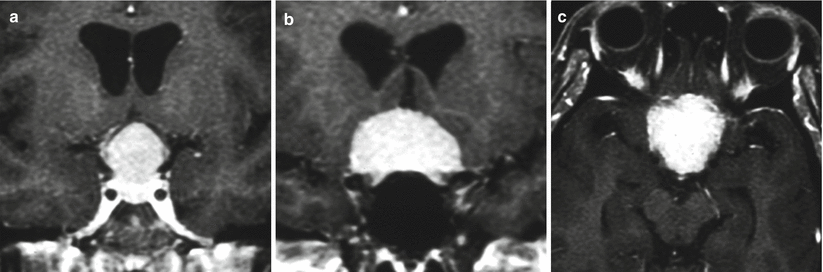
Fig. 28.2
Meningioma. (a, b) Coronal T1-weighted gadolinium-enhanced images. (c) Axial T1-weighted gadolinium-enhanced image. There is an enhancing mass centered in the planum sphenoidale extending to the anterior sella
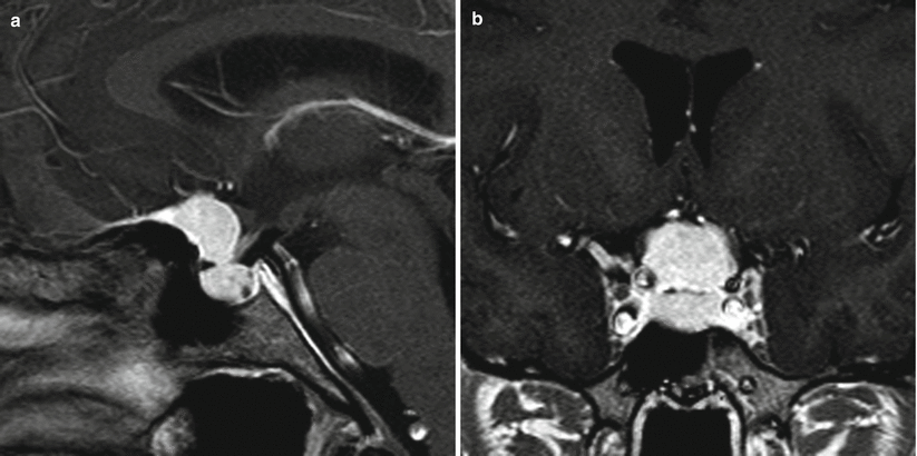
Fig. 28.3
Tuberculum sellae meningioma. (a) Sagittal T1-weighted gadolinium-enhanced image. (b) Coronal T1-weighted gadolinium-enhanced image. A dural-based enhancing mass is centered in the planum sphenoidale, extending to the anterior sella. The mass is above the pituitary gland and anterior to the pituitary stalk. The mass abuts bilateral cavernous internal carotid arteries without encasement
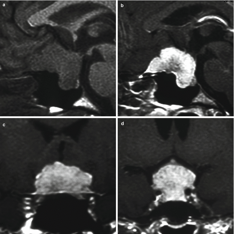
Fig. 28.4
Meningioma. (a) Sagittal T1-weighted precontrast image. (b) Sagittal T1-weighted gadolinium-enhanced image. (c, d) Coronal T1-weighted gadolinium-enhanced images. A dural-based enhancing mass is centered in the planum sphenoidale, extending to the anterior sella. The mass is above the pituitary gland, which is flattened against the sellar floor. The mass surrounds bilateral cavernous internal carotid arteries
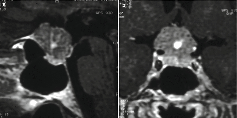
Fig. 28.5
Meningioma. (a) Sagittal T1-weighted gadolinium-enhanced image. (b) Coronal T1-weighted gadolinium-enhanced image. A heterogeneously enhancing mass is located in the suprasellar region, with a dural tail in the planum sphenoidale. The mass is above the pituitary gland. The mass surrounds the right cavernous internal carotid artery
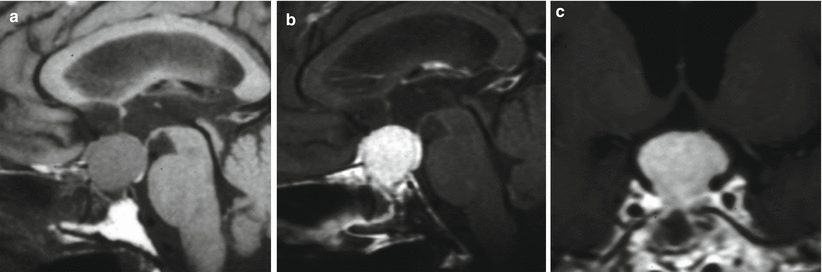
Fig. 28.6
Meningioma. (a) Sagittal T1-weighted precontrast image. (b) Sagittal T1-weighted gadolinium-enhanced image. (c) Coronal T1-weighted gadolinium-enhanced image. There is an enhancing mass centered in the suprasellar region, with a dural tail in the planum sphenoidale. The pituitary gland is not well visualized. The mass abuts the cavernous internal carotid arteries bilaterally
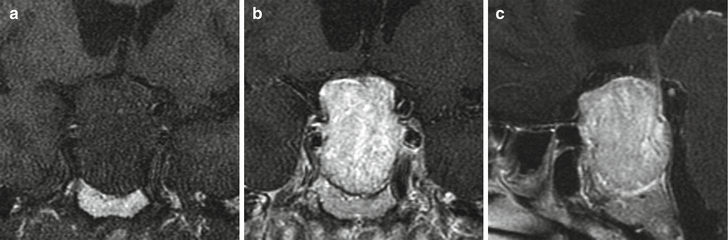
Fig. 28.7
Meningioma. (a) Coronal T1-weighted precontrast image. (b) Coronal T1-weighted gadolinium-enhanced image. (c) Sagittal T1-weighted gadolinium-enhanced image. A heterogeneously enhancing mass is centered in the sella and the suprasellar regions. The pituitary gland is not well visualized, and the sellar floor appears eroded. Superiorly, the mass elevates the optic chiasm
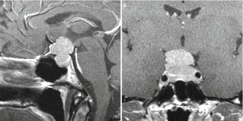
Fig. 28.8
Diaphragma of sellae meningioma. (a) Sagittal T1-weighted gadolinium-enhanced image. (b) Coronal T1-weighted gadolinium-enhanced image. An enhancing mass is centered in the sella/suprasellar region. There is a small dural attachment at the planum sphenoidale. The pituitary gland is not well visualized. The mass surrounds the right cavernous internal carotid artery
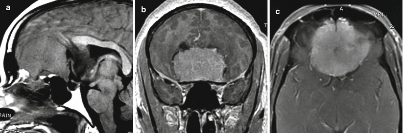
Fig. 28.9
Meningioma. (a) Sagittal T1-weighted precontrast image. (b) Coronal T1-weighted gadolinium-enhanced image. (c) Axial T1-weighted gadolinium-enhanced image. There is a large, enhancing mass arising from the olfactory groove and planum sphenoidale, compressing the frontal lobes. The pituitary gland is separate from the mass
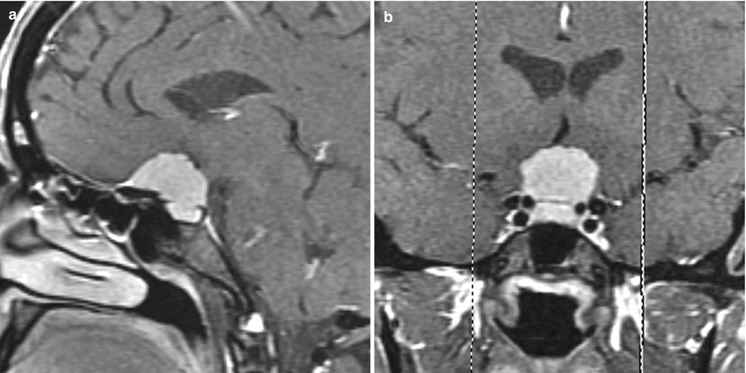
Fig. 28.10
Meningioma. (a) Sagittal T1-weighted gadolinium-enhanced image. (b) Coronal T1-weighted gadolinium-enhanced image. An enhancing mass in the planum sphenoidale extends to the anterior sella. The pituitary gland is seen in the inferior aspect of the sella, immediately inferior to the mass
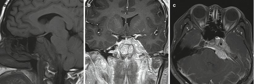
Fig. 28.11
Meningioma. (a) Sagittal T1-weighted precontrast image. (b) Coronal T1-weighted gadolinium-enhanced image. (c) Axial T1-weighted gadolinium-enhanced image with fat saturation. An enhancing mass is seen in the left sphenoid-clival ligament, with components in both left middle cranial fossae involving the cavernous sinus, as well as in the prepontine region, mildly effacing the pons. The mass likely encases the left cavernous internal carotid artery, but flow void (c) indicates that it remains patent
28.3 Histopathology
Meningiomas are typically benign lesions with meningothelial, fibroblastic, or mixed components. Meningothelial whorls and psammoma bodies are common (Fig. 28.12).
The World Health Organization (WHO) classifies meningiomas as benign (WHO I), atypical (WHO II), or anaplastic/malignant (WHO III). Atypical and anaplastic meningiomas are aggressive subtypes with a higher MIB-1 labeling index, mitotic features, and nuclear pleomorphism.
WHO Grade II meningiomas are characterized by any of these parameters:
A mitotic index of 4 or more per 10 high-power field
At least three of the five following parameters:
Sheeting architecture (loss of whorling and/or fascicles)
Small cell formation (high nucleus-to-cytoplasm ratio)
Macronucleoli
Hypercellularity
Spontaneous necrosis (i.e., not induced by embolization or radiation)
Brain invasion
WHO Grade III meningiomas are characterized by a mitotic index of 20 or more per high-power field and/or frank anaplasia (sarcoma-, carcinoma-, or melanoma-like histology).
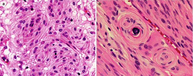
Fig. 28.12
Meningioma. Similar to meningiomas arising in any portion of the CNS, sellar tumors may exhibit a variety of meningioma subtypes. (a) A meningothelial meningioma shows well-differentiated cells with prominent syncytial arrangements; note the characteristic intranuclear pseudoinclusion. (b) A transitional meningioma is composed of elongated spindle cells arranged in fascicles side-by-side with well-formed meningothelial whorls and psammoma bodies
28.4 Clinical and Surgical Management
The surgical approach is frequently dictated by the relationship of the tumor to the neurovascular structures of the sellar and parasellar region, particularly the optic nerves and chiasm, carotid arteries, and pituitary gland and stalk. The position of the optic chiasm relative to the tuberculum sellae (pre-fixed or post-fixed optic chiasm) often plays a major role in the selection of the approach [11].
Microscopic analysis has shown that meningiomas frequently demonstrate evidence of microinvasion of the cranial nerves and carotid artery wall, making a gross total resection unsafe (if not impossible) in some cases [12, 13].
The main goals of surgery are complete tumor resection (when possible), surgical cytoreduction, preservation of visual function, and preservation of pituitary function.Related posts:

Stay updated, free articles. Join our Telegram channel

Full access? Get Clinical Tree


