Fig. 66.1
Encephalocele. Sagittal T1-weighted MR image (a) and sagittal T2-weighted MR image (b) showing a basal encephalocele extending into the nasopharynx (From Chen et al. [10]; with permission)

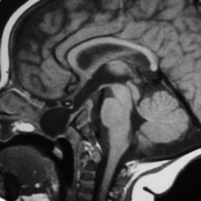
Fig. 66.2
Meningocele. Sagittal T1-weighted MRI showing a transsphenoidal meningocele extending to the posterior portion of the palate (From Franco et al. [11]; with permission)

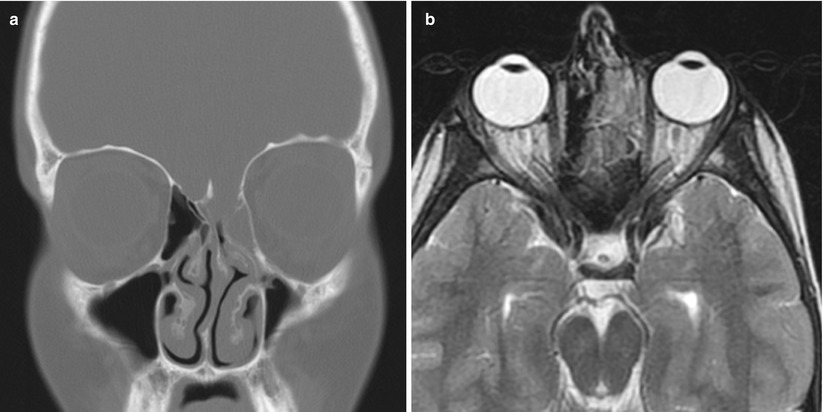
Fig. 66.3
Encephalocele. (a) Coronal CT image. (b) Axial T2-weighted MR image. Soft tissue is present in the left ethmoid (b) and there is a defect in the roof of the left ethmoid (a), consistent with an ethmoid encephalocele

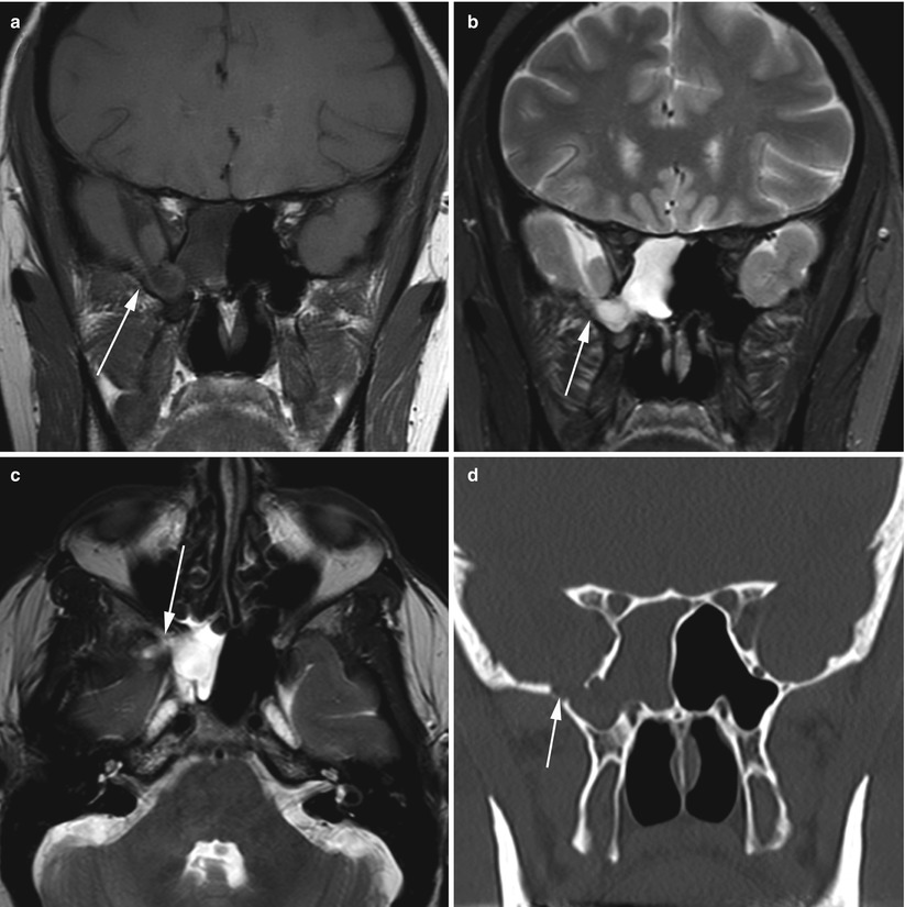
Fig. 66.4
Meningoencephalocele. Coronal T1-weighted MRI (a), T2-weighted MRI (b), and axial T2-weighted MRI (c) showing a large right middle fossa meningoencephalocele (arrow) with extension into the sphenoid sinus. A coronal CT scan (d) shows the bony defect in the floor of the right middle fossa (arrow)

Fig. 66.5
Meningoencephalocele. Sagittal (a) and coronal (b) T1-weighted MR images in a patient with a middle fossa meningoencephalocele extending into the sphenoid sinus. An axial T2-weighted MR image (c) shows a hyperintense CSF collection arising from the left temporal region and extending into the sphenoid sinus
66.3 Histopathology
Marked gliosis of the brain is often noted in an encephalocele. Evidence of lymphocytic infiltrate and dural thickening may be present in cases of infection.
66.4 Clinical and Surgical Management
Both open cranial and endoscopic endonasal approaches have been widely utilized for the surgical repair of symptomatic meningoencephaloceles. Rates of successful repair appear similar for both approaches, but complication rates may be lower in patients treated with endoscopic surgery [14].
In recent years, extended endonasal endoscopic approaches have been used to repair the skull base defects. These cases often involve endonasal transmaxillary and/or transpterygoid approaches; the use of a pedicled nasoseptal flap and possibly a lumbar drain is recommended for larger defects. In many cases, the encephalocele brain is nonfunctional (or even a seizure focus) and should be resected [3, 15].
Outcomes following repair of sphenoid meningoencephaloceles are excellent, with high success rates (>90 %) and rare recurrences [3, 16–18].
In rare cases of midline transsellar encephaloceles, thorough screening for hypopituitarism should be performed, with hormone replacement.
References
1.
Lee TJ, Chang PH, Huang CC, Chuang CC. Endoscopic treatment of traumatic basal encephaloceles: a report of 8 cases. J Neurosurg. 2008;108:729–35.CrossRefPubMed
Stay updated, free articles. Join our Telegram channel

Full access? Get Clinical Tree


