Fig. 50.1
Metastasis from breast cancer. (a) Sagittal T1-weighted gadolinium-enhanced MR image. (b) Coronal T1-weighted gadolinium-enhanced image. A large, heterogeneously enhancing mass is centered in the sella with suprasellar extension. The mass abuts the cavernous internal carotid arteries (ICAs) bilaterally. The normal pituitary gland cannot be distinguished from the mass
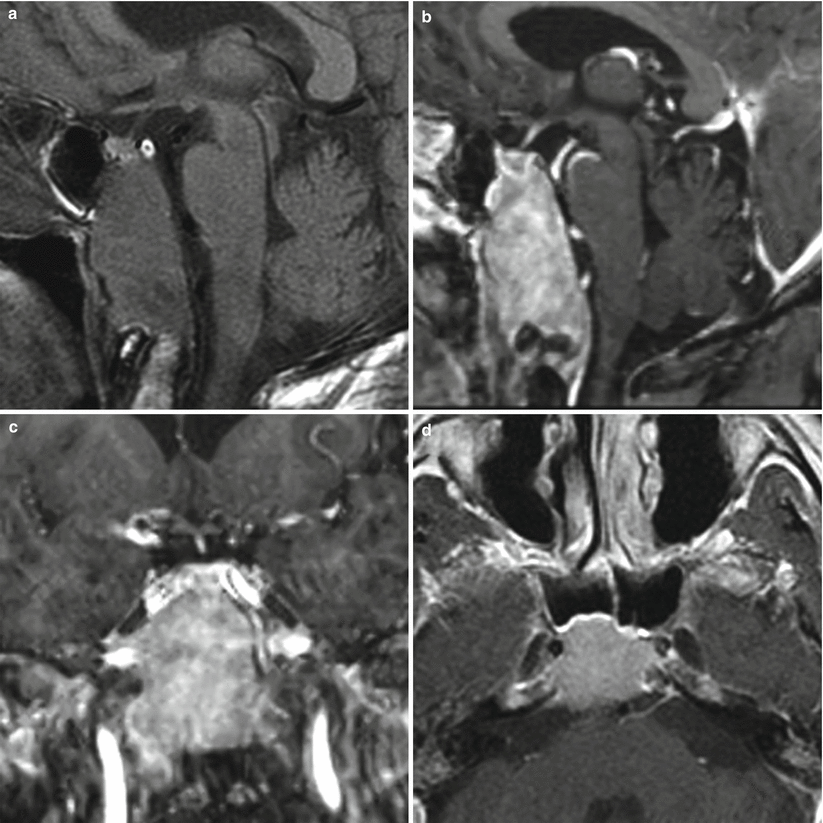
Fig. 50.2
Metastasis. (a) Sagittal T1-weighted precontrast MR image. (b) Sagittal T1-weighted gadolinium-enhanced image. (c) Coronal T1-weighted gadolinium-enhanced image. (d) Axial T1-weighted gadolinium-enhanced image. There is a large, heterogeneously enhancing mass replacing most of the clivus. The mass abuts the cavernous ICAs bilaterally. The normal pituitary gland is seen within the sella, above the mass
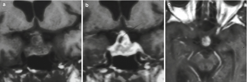
Fig. 50.3
Metastasis. (a) Coronal T1-weighted precontrast MR image. (b) Coronal T1-weighted gadolinium-enhanced image. (c) Axial T1-weighted gadolinium-enhanced image. There is an enhancing mass in the sella, with suprasellar extension abutting the optic chiasm. The mass abuts the cavernous ICAs bilaterally. The normal pituitary gland cannot be distinguished from the mass
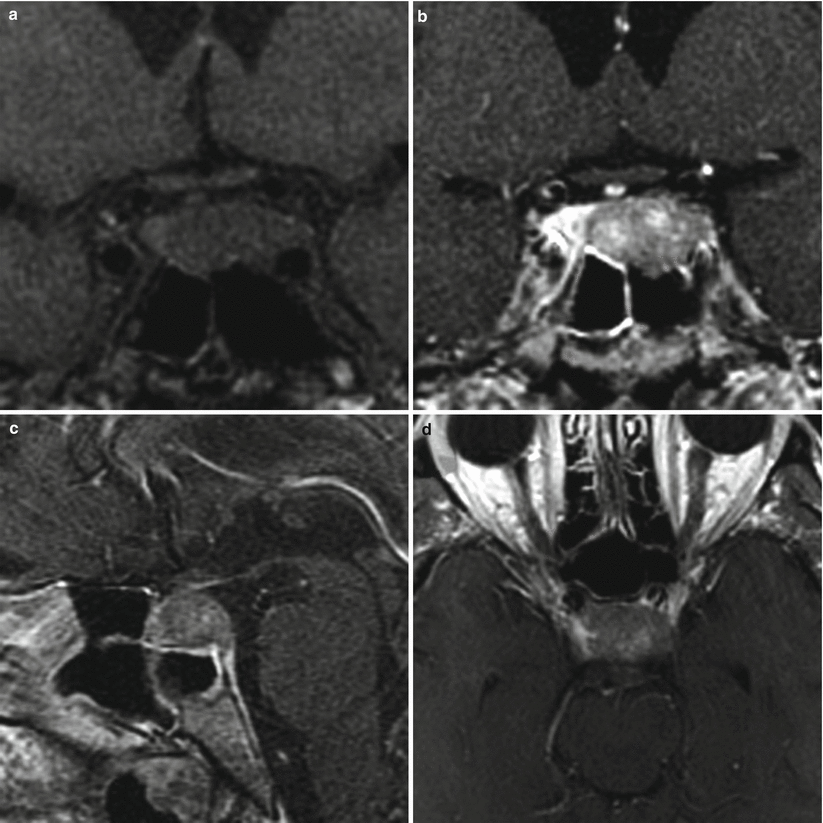
Fig. 50.4
Metastasis. (a) Coronal T1-weighted precontrast MR image. (b) Coronal T1-weighted gadolinium-enhanced image. (c) Sagittal T1-weighted gadolinium-enhanced image. (d) Axial T1-weighted gadolinium-enhanced image. An enhancing mass in the left aspect of the sella abuts the left cavernous ICA. The normal pituitary gland is to the right of the mass
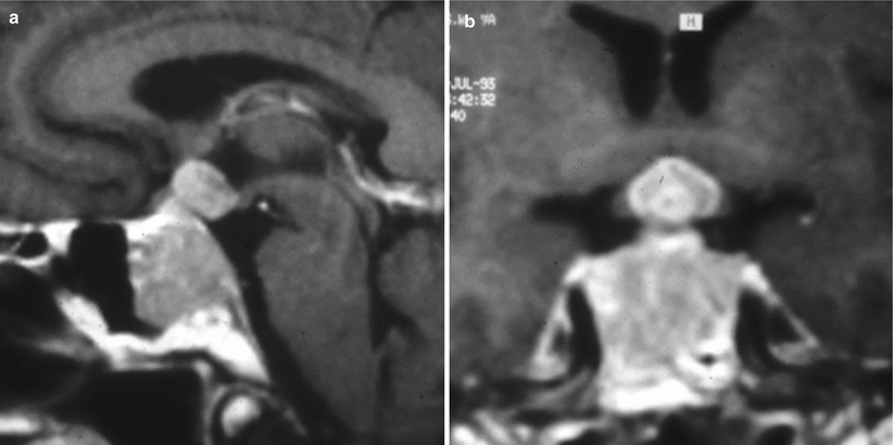
Fig. 50.5
Metastasis. (a) Sagittal T1-weighted gadolinium-enhanced MR image. (b) Coronal T1-weighted gadolinium-enhanced image. There is an enhancing mass replacing the superior clivus, extending to the sella and the anterior floor of the third ventricle. The normal pituitary gland cannot be distinguished from the mass
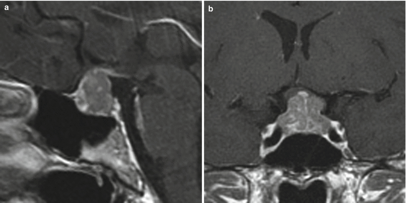
Fig. 50.6
Metastasis from breast cancer. (a) Sagittal T1-weighted gadolinium-enhanced MR image. (b) Coronal T1-weighted gadolinium-enhanced image. A sellar/suprasellar enhancing mass displaces the optic chiasm superiorly and abuts the cavernous ICAs bilaterally. The normal pituitary gland cannot be distinguished from the mass
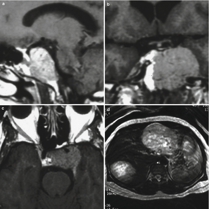
Fig. 50.7
Metastasis from pheochromocytoma. (a) Sagittal T1-weighted gadolinium-enhanced MR image. (b) Coronal T1-weighted gadolinium-enhanced image. (c) Axial T1-weighted gadolinium-enhanced image. (d) Axial T1-weighted, gadolinium-enhanced image showing a heterogeneously enhancing left adrenal mass. An enhancing mass is centered in the upper clivus, involving the sella and extending to the left cavernous sinus and encasing the left ICA (c). The normal pituitary gland cannot be distinguished from the mass
50.3 Histopathology
Metastatic tumors to the pituitary gland have been reported to spread from many primary cancers, including breast, lung, gastrointestinal, prostate, renal, melanoma, and thyroid cancers [6].
In some cases, it may be difficult to differentiate metastatic tumors from pituitary adenomas. Special immunomarkers, such as thyroid transcription factor 1, cdx-2, or peptides of gut or lung origin, can be used to screen for a primary source [14].
Primary carcinomas metastasize to various regions [3, 13]:
Posterior pituitary gland alone (57 %)
Anterior pituitary gland alone (13 %)Related posts:

Stay updated, free articles. Join our Telegram channel

Full access? Get Clinical Tree


