• Lateral fluoroscopic visualization is essential to confirm the appropriate level.
• The incision is typically made 4 cm lateral to the midline. The tube is docked onto the lateral aspect of the pars interarticularis and the inferior transverse process.
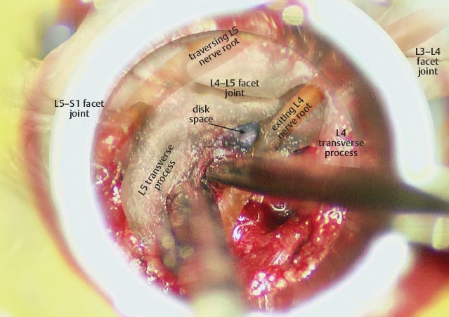
• The tube is docked lateral to the L4–L5 facet joint.
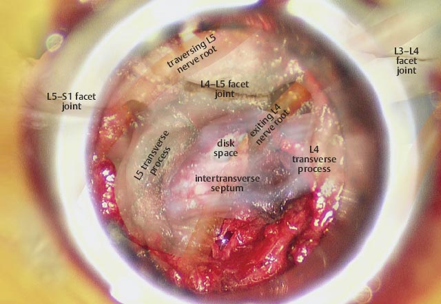
• The intertransverse septum is exposed and is detached from the inferior transverse process with a curved curette.
Related posts:


Stay updated, free articles. Join our Telegram channel

Full access? Get Clinical Tree


