• A lateral fluoroscopic image is used to identify the level in question.
• An incision is made 0.5 cm lateral to the midline.
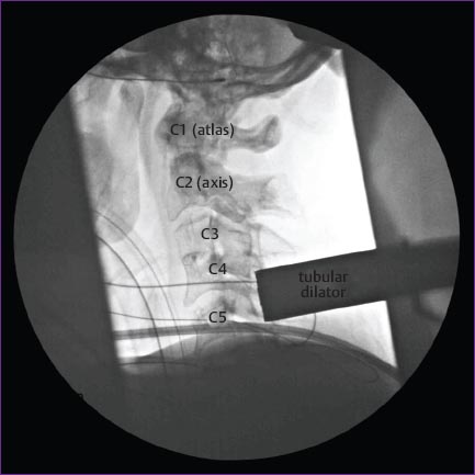
• Tubular dilators (18 mm) are used to spread the paraspinal muscles.
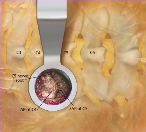
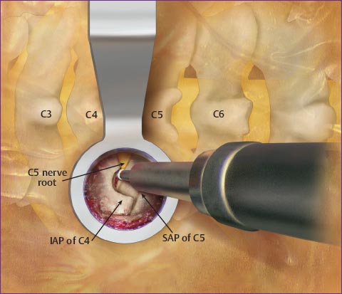
• The soft tissue is removed, exposing the medial half of the facet joint and the inferior portion of the superior lamina.
Related posts:


Stay updated, free articles. Join our Telegram channel

Full access? Get Clinical Tree


