Minimally Invasive Techniques for Lumbar Disorders

CHAPTER 307 Minimally Invasive Techniques for Lumbar Disorders


Minimally invasive techniques have evolved during the past decade to affect all aspects of lumbar spine surgery, from congenital disorders to adult deformity. Surgical series have demonstrated efficacy for decompressive procedures,1 short-segment lumbar fusions,2,3 and intradural exposures to spinal tumors47 and to tethered cords.8 More recent efforts have involved applying this experience to reduce the morbidity of deformity procedures. The advantages of minimally invasive spine surgery include decreased postoperative pain, more rapid postoperative mobilization, shorter length of hospitalization, shorter postoperative recovery times, and less disruption to the paraspinal muscles and ligaments that contribute to the maintenance of proper spine biomechanics.


From a technical perspective, several major themes recur throughout the minimally invasive lumbar spine experience. First, muscle dilators permit the introduction and placement of tubular retractors directly over the site of pathology with minimal soft tissue disruption. Second, a hemilaminar approach using a drill with a dural guard enables contralateral exposure sufficient to perform bilateral decompressive operations and intradural tumor surgery. Third, percutaneous pedicle screw systems now permit placement of posterior stabilization to a theoretically unlimited number of levels without paraspinal muscle dissection. Finally, various other advances in instrumentation placement systems and retractor systems are dramatically decreasing the size of the exposures for many procedures.


Lumbar Diskectomy


Various minimally invasive techniques have been described to treat lumbar disk herniations. The most studied and most accepted of these involves a traditional lumbar microdiskectomy performed through a tubular retractor. An operating microscope or an endoscope can be used for visualization depending on surgeon preference. This procedure is distinguished from the so-called percutaneous diskectomy or endoscopic diskectomy, whereby a trocar is directed into the disk space under fluoroscopic guidance, and disk material is accessed and removed from within the anulus with the aid of specialized instruments. Although good surgical results have been reported using the former technique, it has not gained widespread acceptance and remains conceptually difficult to most neurosurgeons.


Minimally invasive lumbar microdiskectomy, as it is known in the neurosurgical community, involves a similar procedure to traditional microdiskectomy but uses muscle dilators and a tubular retractor to access the interlaminar space with less soft tissue damage. It is performed routinely through tubes ranging from 14 to 22 mm in diameter and has been successfully applied to recurrent disk herniations9,10 and far lateral disk herniations,11,12 in addition to standard disk herniations. Originally developed using an endoscope for visualization, many practitioners use the operating microscope to perform the procedure through the same exposure, and excellent results have been reported.13 Whether a simple lumbar diskectomy using a tubular retractor has significant clinical advantage over an open diskectomy with a conventional subperiosteal muscle dissection and muscle retraction with a Taylor or Thompson-Farley retractor remains a matter of debate.14,15 However, when one considers the increased incisional length and muscle dissection required for proper exposure during a midline approach in heavy patients with typical disk herniations or in all patients with far lateral disk herniations, the advantages of using a tubular retractor centered directly over the site of pathology become obvious.


The procedure is performed as follows. Fluoroscopy is used to center the incision over the correct disk space, about 1 cm off of midline. Initially, a K wire is introduced through a stab incision to center the operation over the junction of the lamina and the inferior articular process of the rostral vertebral level. The incision is extended to accommodate the working channel. Progressively larger muscle dilators are passed, and a working channel of the appropriate length and desired diameter is introduced and fixed to a flexible arm. Cauterization of the remaining soft tissue exposes the inferior lamina, medial facet, and interlaminar space. Now, the procedure is performed in the standard fashion, although bayoneted instruments and an angled drill can be helpful to allow an unobstructed view of the operative field. An endoscope permits a superior view of the operative field and a more comfortable operating position but requires habituation to operating in two dimensions.


Stenosis Decompression


Although the advantages of minimally invasive techniques for microdiskectomy may be debatable, the advantages are clearer for patients with lumbar stenosis requiring a bilateral decompression. Through the same-sized incision as a microdiskectomy, a one-level or two-level stenosis decompression can be performed. Several variations of this procedure have been described, but all share the essential strategy of a bilateral decompression through a hemilaminar approach. This strategy has been proved feasible and effective in large clinical series.1,16


The incision is slightly more lateral than for a microdiskectomy, typically 1.5 cm off midline, to optimize the angle for contralateral decompression, but the process of retractor placement and the final retractor position are the same. An osseous foraminotomy is performed leaving the ligamentum flavum intact for dural protection. After an initial ipsilateral decompression is performed, the retractor is redirected contralaterally. Pulling back the working channel a few millimeters can facilitate this redirection. The base of the spinous process is cleaned of soft tissue and drilled away, using a hemilunar sleeve to protect the dura. The contralateral pedicle and foramen are palpated, and the inner table of the lamina and contralateral facet are drilled away. At this point, the ligamentum flavum is removed, and further osseous decompression of the contralateral foramen can be performed with a drill or Kerrison rongeur as necessary. Fluoroscopy can be useful to confirm the extent of contralateral decompression. After the contralateral decompression, the working channel is redirected again and the ipsilateral foraminotomy is completed.


For two-level decompressions, the initial dilation should be performed at the midpoint of the intermediate lamina. Once the working channel is in place, the largest dilator is used to aim the working channel either rostrally or caudally to access the interlaminar space of the first level to be decompressed. After decompression of the first level, the channel is aimed in the other direction for decompression of the second level. Alternatively, the surgeon can perform separate dilations for each level through the same skin incision.


Lumbar Fusion


There are currently three minimally invasive techniques for lumbar fusion. The most widely practiced is the minimally invasive transforaminal interbody fusion (TLIF). The others are the transpsoas lateral interbody fusion, commercially termed XLIF (Nuvasive, San Diego, CA) and DLIF (Medtronic, Memphis, TN), and the transsacral L5-S1 screw, termed AxiaLIF or Trans1. The Trans1 screw is exclusively for L5-S1 fusion and involves a perirectal, transsacral approach to the L5-S1 disk space. The transpsoas, lateral interbody fusion can be applied to all interspaces except L5-S1. The minimally invasive TLIF is the most versatile and can be applied to all levels.


Minimally Invasive Transforaminal Interbody Fusion


The indications for minimally invasive fusions are the same as for open fusions. The best studied of these techniques is the minimally invasive TLIF developed by Foley and Fessler.2,17,18 Its advantages include less blood loss, decreased perioperative narcotic requirements, and decreased length of stay compared with open lumbar fusions. Beyond a clear perioperative superiority, surgical results from open and minimally invasive fusions are similar in terms of validated self-reported patient outcomes after 1 year.3


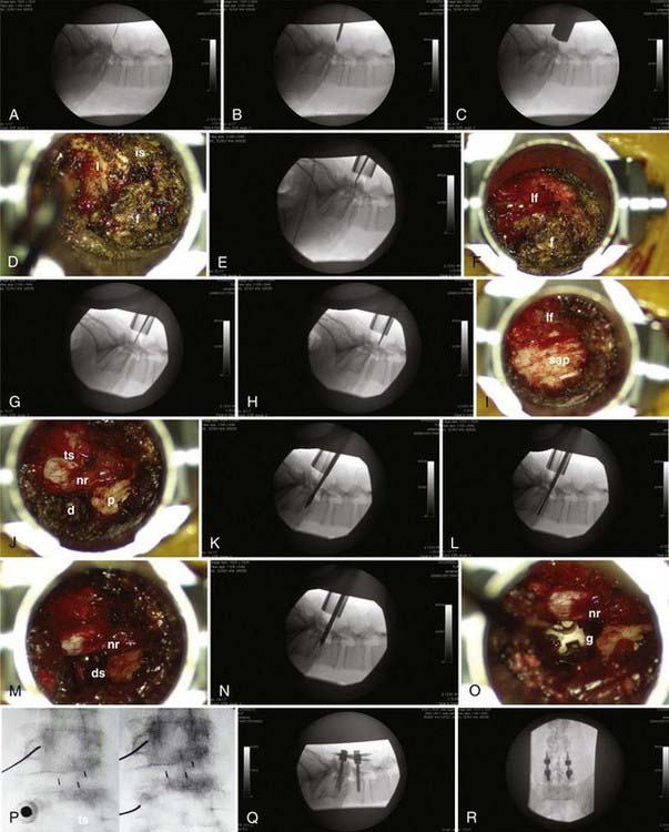
The procedure can be considered in two parts (Fig. 307-1): the interbody fusion and the percutaneous placement of pedicle screw fixation. An incision 2.5 to 3 cm long is made 3 to 5 cm off of midline. This distance is determined by the thickness of overlying soft tissues and can be measured on preoperative imaging to provide the desired working angle. A K wire is angled medially 35 to 45 degrees and docked on the facet joint. After muscle dilation, a working channel 22 to 26 mm is introduced directly over and perpendicular to the disk space, spanning the pedicles. After removal of residual soft tissue, the lamina, facet, and interlaminar space are identified. The canal is defined with curets, and a laminotomy is performed, exposing the ligamentum flavum. The inferior articular process is removed using an osteotome, drill, or rongeurs. All bone is saved for the interbody arthrodesis. The superior articular process is removed, as is the ligamentum flavum. The pedicle inferior to the disk space is identified, as are the exiting nerve root and disk anulus. The anulus is cleaned off of fat and epidural veins and incised. A series of pituitary rongeurs, curets, and scrapers are used to remove the intervertebral disk. An interbody graft is placed along with the arthrodesis material of the surgeon’s choice. Hemostasis is achieved, and the working channel is removed.


image

FIGURE 307-1 Minimally invasive transforaminal interbody fusion. A, The K wire is inserted through a stab incision and docked on the facet orthogonal to the disk space. B and C, Sequential dilators expand the opening to accommodate a 28-mm working port. D, After removal of a thin layer of residual soft tissue, the superficial osseous structures overlying the disk space are revealed (f, facet; is, interlaminar space; l, lamina). E, The plane between the lamina and the interlaminar ligaments is developed with an angled curet, and the level is confirmed. F, A laminotomy is performed, exposing the ligamentum flavum (lf). G-I, Using osteotomes under fluoroscopic guidance, the left inferior articular process of L4 is removed, revealing the left superior articular process (sap) of L5. The ligamentum flavum is removed, along with the facet overlying the disk space and nerve root. J, The thecal sac (ts), nerve root (nr), disk (d), and pedicle (p) are identified, and the epidural vessels over the disk space are cauterized and divided. K-M, A wedge chisel is used to enter the disk space (ds), and a diskectomy is performed using a variety of instruments. N and O, After end-plate preparation and packing of the disk space with rhBMP-2 and local bone, the interbody graft (g) is placed under fluoroscopic guidance. The working channel is removed, and pedicle screws are placed percutaneously. P, A hollow biopsy needle is used to guide trajectories using the bull’s-eye technique, and pedicles are cannulated using guidewires. A cannulated tap and screws permit percutaneous pedicle screw placement, and rods are swung into position through separate incisions using the Sextant system. Q and R, Intraoperative x-rays confirm satisfactory construct position.

Stay updated, free articles. Join our Telegram channel

Aug 7, 2016 | Posted by in NEUROSURGERY | Comments Off on Minimally Invasive Techniques for Lumbar Disorders

Full access? Get Clinical Tree

Get Clinical Tree app for offline access