FIGURE 52.1 Opisthotonos in a patient suffering from tetanus; painting by Sir Charles Bell, 1809. Dr. Bell was a noted artist as well as physician. (see Chapter 16.)
Stiffness and rigidity of the neck may occur in other conditions. A common problem is to distinguish restricted neck motion due to cervical spondylosis or osteoarthritis from nuchal rigidity. Patients with osteoarthritis typically have difficulty with rotation and lateral bending of the neck; these motions are usually preserved in patients who have meningismus, unless the meningeal irritation is extremely severe. Restricted neck motion may also occur with retropharyngeal abscess, cervical lymphadenopathy, neck trauma, and as a nonspecific manifestation in severe systemic infections. Extrapyramidal disorders, particularly progressive supranuclear palsy, may also cause diffuse rigidity of the neck muscles. Meningeal signs may occur with increased spinal fluid pressure, and nuchal rigidity may be a manifestation of cerebellar tonsillar (foramen magnum) herniation. Meningeal irritation may also cause resistance to movement of the legs and back, with the patient lying with his legs drawn up and resisting passive extension.
Kernig’s Sign
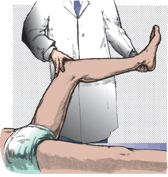
There is some variability in the descriptions of how to elicit a Kernig’s sign. Kernig described an involuntary flexion at the knee when the examiner attempted to flex the hip with the knee extended. The more common method is to flex the hip and knee to right angles and then attempt to passively extend the knee. This movement produces pain, resistance, and inability to fully extend the knee; another definition of Kernig’s sign is inability to extend the knee to over 135 degrees while the hip is flexed (Figure 52.2). There is some overlap between Kernig’s sign and Lasegue’s (straight leg raising) sign. The technique is similar, but Lasegue’s sign is used to check for root irritation in lumbosacral radiculopathy (see Chapter 47). Both the Kernig sign and straight leg raising are positive in meningitis because of diffuse inflammation of the nerve roots and meninges, and positive with acute lumbosacral radiculopathy because of focal inflammation of the affected root. In radiculopathy, the signs are usually unilateral, but in meningitis they are bilateral.

FIGURE 52.2 Method of eliciting Kernig’s sign.
Brudzinski’s Neck Sign
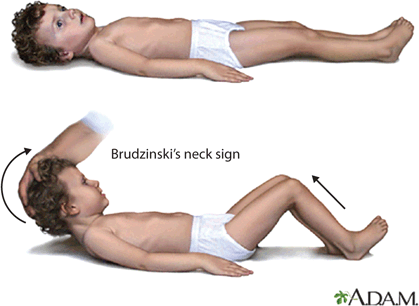
Placing one hand under the patient’s head and flexing the neck while holding down the chest with the other hand causes flexion of the hips and knees bilaterally (Figure 52.3). With severe meningismus, it may not be possible to hold the chest down, and the patient may be pulled into a sitting position with only the examiner’s hand behind the head. Occasionally, there may be extension of the hallux and fanning of the toes, and sometimes arm flexion. The leg may fail to flex on one side when meningeal irritation and hemiplegia coexist.

FIGURE 52.3 Brudzinski’s sign. Flexing the neck causes the knees to flex.
Stay updated, free articles. Join our Telegram channel

Full access? Get Clinical Tree


