7 Central nervous system vascular pathology is uncommon in children, but there are rare forms of vasculopathy unique to this age group, as well as presentations in childhood of some disorders that are more commonly seen in older individuals (Table 7.1). Table 7.1 Arthrogryposis, pterygia, muscular hypoplasia, and pulmonary hypoplasia are found at necropsy (Fig. 7.1a). Bubble-like hemispheres that are macroscopically indistinguishable from those seen in encephaloclastic hydranencephaly have a diaphanous pallium studded with calcifications (Fig. 7.1b). 7.1 Proliferative vasculopathy and hydranencephaly–hydrocephaly in a 17-week fetus. The thin, disorganized pallium has a narrow immature cortical plate. Glomeruloid endothelial vasculopathy, which is unique to this disorder, is prominent throughout the CNS (Fig. 7.2). Endothelial cells in the glomeruloid structures have PAS-positive intracytoplasmic inclusions, which appear on electron microscopy as homogeneous granular material surrounded by rough endoplasmic reticulum (Fig. 7.3). 7.2 Proliferative vasculopathy and hydranencephaly–hydrocephaly in a fetus. 7.3 Proliferative vasculopathy and hydranencephaly–hydrocephaly. This sporadic disorder causes stillbirth or premature delivery with minimal survival. There is diffuse angiodysplasia, consisting of ectatic capillaries and veins. These are present in the leptomeninges, cerebral cortex, cerebellum, and brain stem. The angiodysplasia is complicated by thrombosis, infarction, hemorrhage, hemosiderin-laden macrophages, gliosis and calcification in the cerebral cortex and white matter (Fig. 7.4). 7.4 Meningocerebral angiodysplasia and renal agenesis. The vascular malformation is fed by one or both of the posterior cerebral arteries or their branches resulting in dilatation of the vein of Galen and enlargement of the cerebral venous system and sinuses (Fig. 7.5). The enlarging vascular mass may compress the aqueduct causing hydrocephalus, or may undergo calcification or thrombosis. Microscopically, veins show the typical changes of ‘arterialization’, e.g. hyperplasia and hypertrophy of the intima and muscularis, that result from increased intraluminal pressure. 7.5 Vein of Galen aneurysm (arteriovenous fistula). Lymphocytic infiltration of the media and destruction of the elastica of affected vessels are associated with giant cells and followed by fibrosis and thickening. The blood vessels are further narrowed by superimposed intimal proliferation and atheroma. Thromboembolic disease leads to cerebral ischemia. Only rarely does the arteritic process directly involve cerebral vessels (Fig. 7.6). 7.6 Takayasu’s arteritis in a 14-year-old girl. Hyaline eosinophilic platelet thrombi occlude small arteries and arterioles, which also show striking endothelial swelling. Multiple microinfarcts are present in cerebral and cerebellar gray matter (Fig. 7.8). 7.8 Hemolytic–uremic syndrome.
Miscellaneous pediatric disorders
VASCULAR DISEASES
Fetal presentation
Postnatal onset
Vascular malformation
Proliferative vasculopathy and hydranencephaly–hydrocephaly
Meningocerebral angiodysplasia and renal agenesis
Aneurysm of great vein of Galen
Arteriovenous malformation
Cavernous angioma
Aneurysm
Saccular
Fusiform (basilar)
Infectious (mycotic)
Dissecting
Vasculitis
Kawasaki disease
Takayasu’s arteritis
Coagulopathy
Hemolytic–uremic syndrome
Hemorrhagic shock and encephalopathy syndrome
PROLIFERATIVE VASCULOPATHY AND HYDRANENCEPHALY–Hydrocephaly (Fowler Syndrome)
MACROSCOPIC APPEARANCES
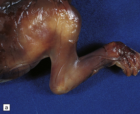
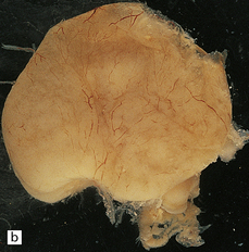
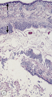
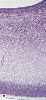
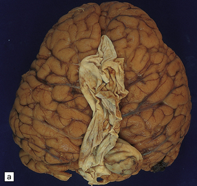
(a) Arthrogryposis and webbing of the elbow joint. (b) Lateral aspect of the cystic cerebral hemisphere demonstrated under water. (c) The extremely thin pallium (between arrows) is barely one-third of the normal. (d) Normal age-matched control for comparison with (c).
MICROSCOPIC APPEARANCES
Glomeruloid vascular structures are widespread in the brain and spinal cord. (a) Glomeruloid vasculopathy in the marginal zone and cortical plate. (b) Prominent vascular expansion involving the intermediate and subventricular zones. (c) Vasculopathy is also visible in the germinal eminence. (d) Glomeruloid structure in the cerebellum (reticulin). (e) Glomeruloid vascular expansion in the anterior horn of the spinal cord.
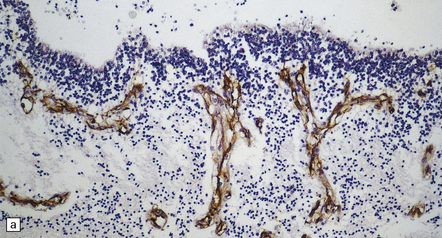
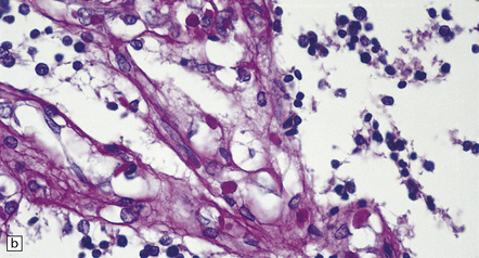
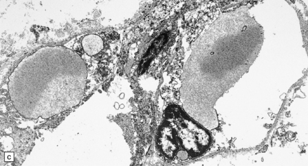
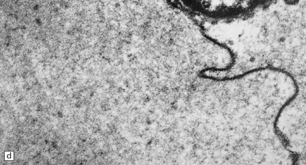
(a) The glomeruloid vascular structures have many small lumina lined by endothelial cells, as demonstrated by lectin histochemistry with Ulex europaeus agglutinin. (b) Some endothelial cells contain PAS-positive intracytoplasmic inclusions. (c) Electron microscopy reveals endothelial inclusions of very varied size comprising moderately electron-dense granular material surrounded by rough endoplasmic reticulum. (d) Close-up of the surrounding rough endoplasmic reticulum.
MENINGOCEREBRAL ANGIODYSPLASIA AND RENAL AGENESIS
MICROSCOPIC APPEARANCES
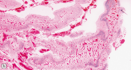
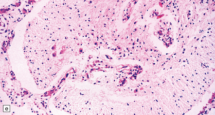
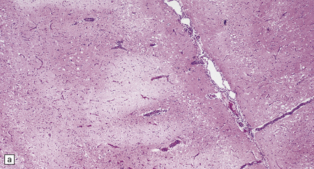
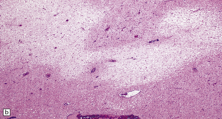
(a) Engorged and markedly ectatic thin-walled vessels are seen within the pia–arachnoid and also in the underlying partially necrotic cerebral parenchyma. Note also the vascular thrombosis and focal calcification. (b) A similar process also affects the cerebellum.
ANEURYSM OF THE VEIN OF GALEN
MACROSCOPIC AND MICROSCOPIC APPEARANCES
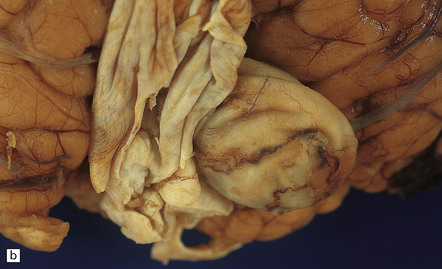
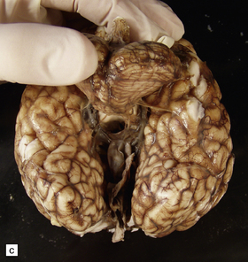
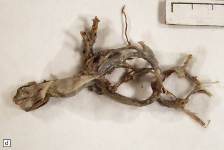
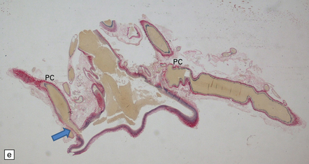
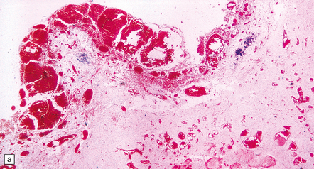
(a,b) The patient is a 15-month-old child who died secondary to extensive encephalomalacia produced by a ‘steal’ of blood from normal brain through the shunt. (c,d) This aneurysm is fed by both posterior cerebral arteries, the right is duplicated. (e) Microscopic section of the aneurysm and its feeding posterior cerebral arteries (PC) with loss of elastic at the fistulous connexion (arrow).
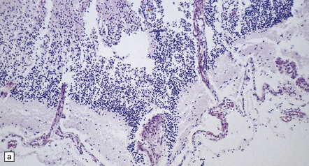
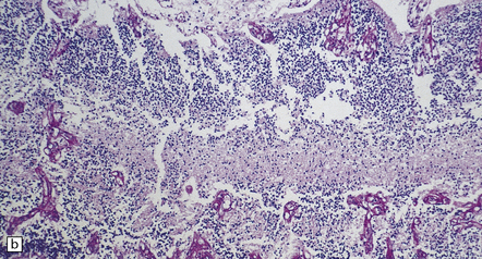
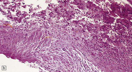
TAKAYASU’S ARTERITIS
MACROSCOPIC AND MICROSCOPIC APPEARANCES
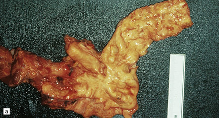
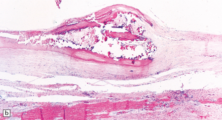
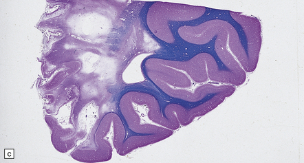
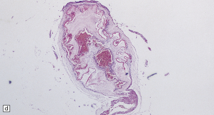
(a) Severe complicated atheroma in the aorta. (b) Histologically, the atheroma consists of calcified and atheromatous thickening of the intima and a mononuclear infiltrate disrupting the media. (c) Old infarction centered on the parieto-occipital vascular boundary zone. (d) All the vessels of the circle of Willis were affected by a chronic arteritic process with intimal thickening, elastic fragmentation, medial atrophy and mononuclear infiltration. The middle cerebral arteries were occluded and recanalized.
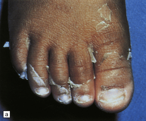
KAWASAKI DISEASE (MUCOCUTANEOUS LYMPH NODE SYNDROME)
 CLINICAL ASPECTS OF KAWASAKI DISEASE
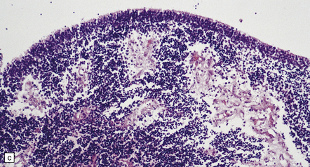
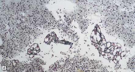
HEMOLYTIC–UREMIC SYNDROME
MACROSCOPIC AND MICROSCOPIC APPEARANCES
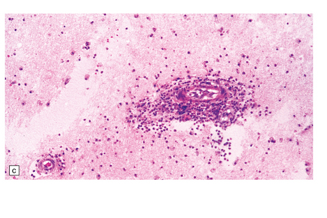
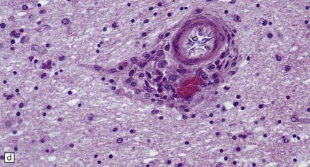
(a) Focal cerebral cortical infarction. (b) Focal cerebellar white matter infarct. (c) Occlusive fibrin thrombus in a small arteriole. (d) Swollen endothelial cells bulging into the lumen of a small vessel.![]()
Stay updated, free articles. Join our Telegram channel

Full access? Get Clinical Tree


Miscellaneous pediatric disorders
 PATHOGENESIS OF FOWLER SYNDROME


 CLINICAL ASPECTS OF TAKAYASU’S ARTERITIS


Only gold members can continue reading. Log In or Register to continue


