Mitochondrial Energy Metabolism Disorders


Mitochondrial Energy Metabolism Disorders


Patricia L. Musolino

Katherine B. Sims


INTRODUCTION

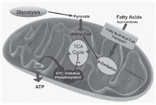
Mitochondrial oxidative-phosphorylation diseases are a clinically heterogeneous group of disorders that arise primarily as a result of dysfunction of the mitochondrial respiratory chain (electron transport chain [ETC]) and can present at any age. More than 70 different polypeptides interact on the inner mitochondrial membrane to form the respiratory chain. The vast majority of subunits are synthesized within the cytosol from nuclear DNA (nDNA) transcripts, but 13 essential subunits are encoded by the 16.5-kb mitochondrial DNA (mtDNA). See Figure 8.1.


CLINCAL PRESENTATION

Because of the presence of mitochondrial heteroplasmy (different amounts of mutant mitochondria) in different cells and tissues, some mitochondrial disorders may affect a single organ but more commonly involve multiple organ systems. Many disorders present with prominent neurologic (central, peripheral, and autonomic) as well as myopathic features. Moreover, variant mtDNA and nDNA mutations can present with the same clinical syndrome, or a single mutation can lead to different combinations of signs and symptoms. Common clinical features of mitochondrial disease include encephalopathy, seizures, dementia, migraine, stroke-like episodes,
ataxia, spasticity, ptosis, external ophthalmoplegia, myopathy (often with exercise intolerance), sensorineural hearing loss/deafness, optic atrophy/pigmentary retinopathy, and nonneurological symptoms (including cardiomyopathy, failure to thrive, hepatic failure, renal tubular acidosis, diabetes mellitus and other endocrinopathies, bone marrow failure, and immunologic alterations (Table 8.1). A high incidence of mid- and late pregnancy loss is a common occurrence that often goes unrecognized. Table 8.2 summarizes “red-flag” clinical S/S that should prompt the primary care team to trigger a mitochondrial disorder diagnostic work-up.


FIGURE 8.1 Simplified Energy Metabolic Pathways Affected in Mitochondrial Disorders.


TABLE 8.1 Signs and Symptoms Suggestive of Mitochondrial Disorder


Neurologic


Cerebral stroke-like lesions in a nonvascular pattern


Basal ganglia dysfunction or mineralization


Encephalopathy: recurrent or w/ low/moderate dosing of valproate


Neurodegeneration


Epilepsia partialis continua, generalized or myoclonic seizures


Myoclonus


Ataxia


MRI findings consistent w/ Leigh disease


Characteristic MRS peaks (lactate peak at 1.3 ppm, TE at 35 & 135; succinate peak at 2.4 ppm)


Cardiovascular


Hypertrophic cardiomyopathy w/ rhythm disturbance


Unexplained heart block in a child


Cardiomyopathy w/ lactic acidosis


Dilated cardiomyopathy w/ muscle weakness


Wolff-Parkinson-White arrhythmia


Ophthalmologic


Retinal degeneration w/ signs of night blindness, colorvision deficits, decreased visual acuity, or pigmentary retinopathy


Ophthalmoplegia/paresis


Fluctuating, dysconjugate eye movements


Ptosis


Sudden- or insidious-onset optic neuropathy/atrophy


Gastroenterologic


Unexplained or valproate-induced liver failure


Severe dysmotility


Pseudo-obstructive episodes


Other


A newborn, infant, or young child w/ unexplained hypotonia, weakness, failure to thrive, and metabolic acidosis (particularly lactic acidosis)


Exercise intolerance that is not in proportion to weakness


Hypersensitivity to general anesthesia


Episodes of acute rhabdomyolysis


Short stature


Autonomic dysfunction/postural orthostatic tachycardia syndrome (POTS)


Adapted from Haas RH, et al. Mitochondrial disease: a practical approach for primary care physicians. Pediatrics. 2007;120:1326-1333.10


DIAGNOSTIC APPROACH

In some individuals, the clinical picture is characteristic of a specific mitochondrial syndrome (e.g., LHON, NARP; see Table 8.2), and the diagnosis can be confirmed by molecular genetic testing of DNA extracted from a blood
sample. In many individuals, such is not the case, and a more structured approach is needed: family history, metabolic screening in blood, urine, and CSF, neuroimaging, cardiac evaluation, and muscle (or other tissue) biopsy for histologic, histochemical, or electron-microscopic evidence of mitochondrial disease, as well as ETC analysis or polography/ATP production (freshly frozen biopsy samples only). Molecular genetic testing for mtDNA and nDNA mutations is often best done in tissue. Diagnostic criteria have been developed1,2 (Table 8.3). Although ability to rule out diagnosis is limited, a proposed algorithm for initial diagnostic work-up is summarized in Table 8.4. In terms of genetic work-up, the strategy is based on clinical constellation, likelihood of mtDNA mutation (clinical syndrome, maternal inheritance), availability of tissue for depletion analysis (muscle, liver), and evidence from biochemical testing.


TABLE 8.2 Clinical Syndromes of Mitochondrial Diseases


Disorder


Primary Features


Additional Features


NUCLEAR DNA MUTATIONS


Alpers-Huttenlocher syndrome


Hypotonia


Seizures


Liver failure


Renal tubulopathy


Leigh syndrome (LS)


Subacute relapsing encephalopathy


Cerebellar, brainstem signs


Infantile onset


Basal ganglia lucencies


Maternal lineage history of neurologic disease, Leigh syndrome, or increased spontaneous abortions


Infantile myopathy and lactic acidosis (fatal and nonfatal forms)


Hypotonia in 1st y of life


Feeding and respiratory difficulties


Fatal form may be associated with a cardiomyopathy and/or de Toni-Fanconi-Debre syndrome


MITOCHONDRIAL DNA MUTATIONS


Progressive external ophthalmoplegia (adPEO and arPEO)


External ophthalmoplegia


Bilateral ptosis


Mild proximal myopathy


Compatible with a normal life span


Kearns-sayre syndrome (KSS)


PEO onset at age <20 y


Pigmentary retinopathy


One of the following: CSF protein >1 g/L Cerebellar ataxia Heart block


Bilateral deafness


Myopathy


Dysphagia


Diabetes mellitus


Hypoparathyroidism


Dementia


Neurogenic weakness with ataxia and retinitis pigmentosa (NARP)


Late-childhood or adultonset peripheral neuropathy


Ataxia


Pigmentary retinopathy


Basal ganglia lucencies


Abnormal electroretinogram


Sensorimotor neuropathy


Myoclonic epilepsy with ragged-red fibers (MERRF)


Myoclonus


Seizures


Cerebellar ataxia


Myopathy


Dementia


Optic atrophy


Bilateral deafness


Peripheral neuropathy


Spasticity


Generalized lipomatosis


Leber hereditary optic neuropathy (LHON)


Subacute painless b/l visual failure


M:F, 4:1


Median age of onset 24 y


Dystonia


Cardiac preexcitation syndromes


Mitochondrial encephalomyopathy with lactic acidosis and stroke-like episodes (MELAS)


Stroke-like episodes at age <40 y


Seizures and/or dementia


Ragged-red fibers and/or lactic acidosis


Diabetes mellitus


Cardiomyopathy (initially hypertrophic, later dilated)


Bilateral deafness


Pigmentary retinopathy


Cerebellar ataxia


Pearson syndrome


Sideroblastic anemia of childhood


Pancytopenia


Exocrine pancreatic failure


Renal tubular defects


Myoclonic epilepsy myopathy sensory ataxia (MEMSA or MIRAS)


Myopathy


Seizures


Cerebellar ataxia


Dementia


Peripheral neuropathy


Spasticity


Coenzyme Q10 deficiency


Encephalopathy, myopathy


Seizures, cerebellar ataxia


Cardiomyopathy


Renal failure


Growth retardation


Early infancy LS (severe form)


Late onset: myopathy, ataxia, seizures, mild encephalopathy


Modified from genereviews.org. Chinnery PF. Mitochondrial Disorders Overview. 2000 Jun 8 [Updated 2010 Sep 16]. In: Pagon RA, Adam MP, Bird TD, et al., editors. GeneReviewsTM [Internet]. Seattle (WA): University of Washington, Seattle; 1993-2013. Available from: http://www.ncbi.nlm.nih.gov/books/NBK1224/


GENETIC COUNSELING

Mitochondrial disorders caused by defects of nuclear DNA (nDNA) may be inherited in an autosomal recessive, autosomal dominant, or X-linked manner. mtDNA defects are transmitted by maternal inheritance. mtDNA deletions generally occur de novo and thus cause disease sporadically, with no significant risk to other family members. mtDNA point mutations and duplications may be transmitted down the maternal line. Thus both males and females can be affected by mtDNA pathologic lesions, but a male does not transmit the mtDNA mutation to his offspring. A female harboring a heteroplasmic mtDNA point mutation may transmit a variable amount of mutant mtDNA to her offspring (heteroplasmy), resulting in considerable
intrafamilial as well as interfamilial clinical variability. Prenatal genetic testing, and interpretation of test results for mtDNA disorders, are difficult because of this mtDNA heteroplasmy and should be deferred to the genetic counselor or mitochondrial expert. nDNA-encoded mitochondrial disorders are increasingly recognized. Up to 1,200 nuclear genes, presumably associated with broad mitochondrial dysfunction, are being currently screened in clinical cohorts. The majority of nDNA-associated disorders, identified to date, are those with infantile encephalopathy features. Increasingly, however, late-onset phenotypes are being recognized (myopathy, ataxia, PEO).


TABLE 8.3 Mitochondrial Disorders Diagnostic Criteria


Clinical Signs and Symptoms (max 4 Points)


Muscular Presentation (max 2 Points)


Cns Presentation (max 2 Points)


Multisystem Disease (max 3 Points)


Metabolic and Imaging Studies (max 4 Points)


Pathology (max 4 Points)


Ophthalmoplegia1


Facies myopathica


Exercise intolerance


Muscle weakness


Rhabdomyolysis


Abnormal EMG


Dev delay


Loss of skills


Stroke-like


Migraine


Seizures


Myoclonus


Cortical blindness


Pyramidal signs


Extrapyramidal signs


Brainstem involvement


Hematology


GI tract


Endocrine/growth


Heart


Kidney


Vision


Hearing


Neuropathy


Recurrent/familial


Elevated lactate1


Elevated L/P ratio


Elevated alanine1


Elevated CSF lactate1


Urinary tricabon acid excretion1


Ethylmalonic aciduria


Stroke-like picture/MRI


Leigh syndrome/MRI1


Elevated lactate/MRS


Ragged-red/blue fibers2


COX-negative fibers2


Reduced COX staining2


Reduced SDH staining


SDH-positive blood vessels1


Abnormal mitochondria/EM1


Score 1: mitochondrial disorder unlikely; score 2-4: possible mitochondrial disorder; score 5-7: probable mitochondrial disorder; score 8-12: definite mitochondrial disorder.1 scores 2 points.2 If in a higher %, scores 4 points.


Modified from Morava E, et al. Mitochondrial disease criteria: diagnostic applications in children. Neurology. 2006;67:1823-1826, with permission.


TABLE 8.4 Screening Algorithm for Patients with Suspected Mitochondrial Disorder


For ALL Patients


Family History and Pedigree


Blood


Basic chemistries


CBC, CPK


LFTs, ammonia


Blood lactate, pyruvate (lactate/pyruvate ratio), coenzyme Q10 (WBC, tissue)


Quantitative plasma amino acids


Plasma carnitines (free, total, esterified) and acylcarnitines


Urine


Quantitative urine organic acids


Echocardiogram & EKG


Ophthalmologic examination


Audiology testing


If Neurological Symptoms:


All of above plus:


Brain MRI/MRS

Only gold members can continue reading. Log In or Register to continue

Stay updated, free articles. Join our Telegram channel

Jun 20, 2016 | Posted by in NEUROLOGY | Comments Off on Mitochondrial Energy Metabolism Disorders

Full access? Get Clinical Tree

Get Clinical Tree app for offline access