Monitoring Techniques for Evaluating Suspected Sleep-Disordered Breathing

Chapter 142 Monitoring Techniques for Evaluating Suspected Sleep-Disordered Breathing


Abstract


Assessment of sleep-disordered breathing represents the most common application of laboratory and home sleep studies. Numerous techniques can provide objective measures of breathing, identify breathing abnormalities, and detect physiologic alterations produced by abnormal respiratory events. Recording techniques, measurement criteria, data acquisition systems, operational paradigms, and the setting where data are collected (home or laboratory) can vary significantly. Over the past 3 decades, some common procedures have evolved and become commonplace in clinical sleep laboratories. In 2007, the American Academy of Sleep Medicine (AASM) published guidelines for laboratory-based techniques for recording sleep-related breathing, method of scoring, and terminology as part of their standardized sleep manual. Additional proposals concerning home sleep testing were published subsequently. Recommendations were based on expert opinion, consensus panels, and published evidence. However, some proposals remain controversial and hotly debated.


Evaluating breathing during sleep has become a component of standard medical practice. Diagnostic methods largely reflect AASM recommendations, but local medical resources and research needs also influence specific procedures. The AASM manual does not provide illustrative examples for the spectrum of sleep-related breathing disorders; however, examples are given in Section 13.


Definitions


Abnormal breathing during sleep can take several different forms, arises from differing etiologies, and goes by many names, including sleep apnea, sleep apnea–hypopnea syndrome, sleep-disordered breathing (SBD), sleep-related breathing disorder (SRBD), periodic breathing, Cheyne-Stokes respiration, and hypoventilation (Box 142-1). SRBDs associated with neurologic lesions are described elsewhere in this volume. Some terms distinguish between different types of sleep-related breathing impairments; others are synonymous. SDB events include episodes of apnea, hypopneas, respiratory effort–related arousals, oxygen desaturations, and snore arousals.


Box 142-1 Sleep-Related Breathing Events, Disorder Classifications, and Parameters


CNS, central nervous system.


The criteria for designating a breathing event as an apnea episode is defined fairly consistently. By contrast, definitions of hypopnea vary widely. Part of the difficulty stems from the fact that a hypopnea episode during sleep (or wakefulness) is not intrinsically abnormal—it is merely a shallow breath. However, a hypopnea that occurs during sleep and is associated with significant oxyhemoglobin desaturation or a central nervous system arousal is deemed pathophysiologic. Another difficulty arises because recording techniques for indexing airflow are uncalibrated and thus qualitative. The airflow signal magnitude from thermistors, thermocouples, capnographs, and nasal pressure transducers correlate poorly with tidal volume. Consequently, operational definitions for hypopnea based on a percentage decrease of flow signal use arbitrary cutpoints (upon which there exists considerable disagreement). Similarly, even though oximetry provides quantitative indices, a desaturation event’s pathophysiologic threshold remains a matter of disagreement, with some setting the criteria at 3% and others requiring drops of 4% or more. Finally, respiratory-related and snore-related arousals certainly seem irrefutably abnormal (because they compromise sleep integrity); however, there are issues concerning detection reliability.


Apnea and hypopnea episodes can be further categorized as obstructive, central, or mixed based on the presence or absence of respiratory effort during the entirety or some part of the breathing event.


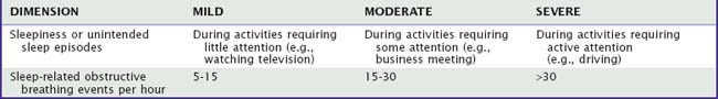
Sleep-disordered breathing severity can be based on a clinical dimension (e.g., sleepiness), event frequency (e.g., number of events per hour), or magnitude of the consequence (e.g., degree of oxyhemoglobin desaturation). Table 142-1 provides examples for dimensionally classifying severity of SDB. There is no general agreement about assigning severity descriptors to indices of SDB; however, two schemes are commonly used. In the first (the liberal) classification, an apnea–hypopnea index (AHI) between 5 and 15 is mild, between 15 and 30 is moderate, and greater than 30 is severe. In the second (the conservative) classification, an AHI between 10 and 20 is mild, between 20 and 50 is moderate, and greater than 50 is severe.1


Methods to Detect Airflow


In general, airflow cessation or near cessation for 10 seconds or more constitutes a sleep apnea episode. Although the American Academy of Sleep Medicine (AASM) manual recommends a thermal sensor to document apnea and a nasal pressure sensor to detect hyponea,2 other methods provide reliable assessment. Occasionally, unidirectional occlusion occurs, with no airflow during attempted inspiration but tiny puffs of expiration. Fully quantitative airflow determination requires pneumotachography. Alternatively, a body box could be used; however, such an approach is unsuitable for sleep studies. Semiquantitative measures are attainable using calibrated inductance plethysmography; however, most clinical evaluations rely on qualitative nasal-oral thermography and nasal pressure to minimize the patient’s discomfort, reduce costs, and simplify acquisition of data. Airflow can also be measured qualitatively by detecting chemical differences between ambient and expired air (e.g., capnography).


Pneumotachography


Several types of pneumotachographs based on different physical principles are in use: differential pressure airflow transducers, ultrasonic flow meters, and hot-wire anemometers. The discussion is limited here to the most widely used technique: the differential pressure flow transducer. In this technique, airflow directed through a cylinder exits through a small resistive field, usually composed of small parallel tubes or a grill promoting laminar flow. The pressure drop across this resistive field is measured using a differential manometer. When flow is laminar, the relationship between the pressure differences and flow is linear. Changes in gas density, viscosity, and temperature alter the pressure–flow relationship. To prevent condensation on the resistive element requires heating; therefore, calibration should be conducted when the pneumotachograph is heated. After correcting for errors introduced by alterations in these physical factors, the flow signal is integrated to determine volume.


Pneumotachography accurately and quantitatively measures airflow volume. The patient usually wears a facemask, the equipment can be bulky and large, and the procedure is usually uncomfortable. Therefore, its routine use for overnight assessment of SDB is problematic. In awake subjects, such invasive devices can increase tidal volume and reduce respiratory rate. Nonetheless, some positive airway pressure machines have built-in pneumotachographs and can be used to monitor airflow and ventilation. Cardiogenic oscillations can also be detected to provide a possible marker for central apnea.3


Thermistors and Thermocouples


Exhaled air is usually warmer than ambient temperature. Air in the lungs is warmed by core body heat, thereby creating a temperature difference between air entering and exiting the respiratory system. Consequently, measuring temperature fluctuation at the nares and in front of the mouth provides a simple surrogate measure of airflow. Measurement is possible using thermistors.


Thermistors are thermally sensitive variable resistors that produce voltage alterations when connected in a low-current (but constant-current) circuit. Low current minimizes the tendency for the thermistor to heat itself. Thermistors maximize sensing area while minimizing sensor size and mass. Small temperature changes can produce large resistance changes that can in turn be transduced with a bridge amplifier. Care must be taken to ensure the thermistor remains below body temperature (i.e., it must not rest on the skin); otherwise, expired air will not be warmed and no resistance change will occur. In such a case, inspiratory activity and a respiratory pause will not be differentiable.


Thermocouples also sense temperature change but use a different approach. Different metals expand at different rates when heated. In a calibrated system, this difference can be transduced to voltage alterations displayable on polygraph systems. Like thermistors, thermocouples are placed in the path of airflow in front of the nares and mouth, where expired air heats the sensor and increases its resistance. The transduced signal reflects oscillation between exhaled warm air and cooled inhaled air, thus providing a trace roughly corresponding to respiratory airflow.


Nasal Airway Pressure


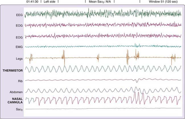
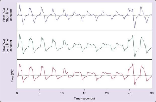
During inspiration, airway pressure is negative relative to atmosphere. By contrast, expiration produces a relatively positive pressure in the airway. The resulting alteration in nasal airway pressure can provide a surrogate estimate of airflow and correlates favorably with pneumotachographically recorded signals.4 The nasal pressure signal also offers greater sensitivity for detecting subtle flow limitations than nasal–oral thermography (Fig. 142-1) when persons breathe through the nose.5 Airflow limitation manifests as pressure trace plateauing during inspiration. A direct current (DC) amplifier provide optimal interface; however, long time-constant alternating current (i.e., a very slowly coupled signal) can suffice. By contrast, rapid coupling can create artifact (Fig. 142-2).


Expired Carbon Dioxide Sensing


Air leaving the lungs has a much higher concentration of CO2 than ambient air. There is always a large CO2 difference between air entering and exiting the respiratory system. Thus, measuring CO2 in front of the nose and mouth can detect expiration, and an infrared analyzer can be used to determine its concentration. Measuring CO2 offers several advantages compared with thermistor, thermocouple, and nasal pressure recordings.


In some patients, the end-of-breath CO2 concentration provides evidence of elevated end-tidal PCO2. The catheters sampling CO2 typically entrain some room air, making the measured CO2 lower than actual end-tidal PCO2. Therefore, an elevated CO2 indicates that true PCO2 is even higher, thereby providing a noninvasive technique (merely sampling the air stream) for detecting hypoventilation.


The shape of the expired CO2 curve can also offer useful information. When a patient’s baseline expired CO2 curve shows a clear-cut plateau, the loss of this plateau (or the curve’s becoming smaller or dome shaped) indicates a change in breathing pattern, usually a reduction in expiratory volume.


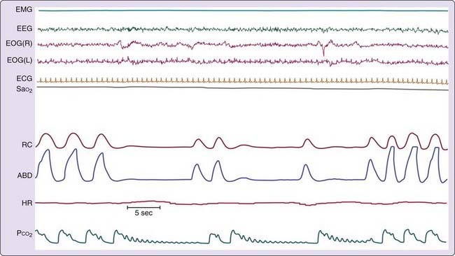
During central apnea, a low-volume catheter system set at its most rapid response time can show cardiogenic oscillations in the CO2 signal. These oscillations result from small volume displacements caused by the beating heart.6 These heartbeat-synchronized oscillations signify a wide-open upper airway (Fig. 142-3).


Infants and children with upper airway obstruction can severely hypoventilate during sleep without observable apnea or hypopnea. Measuring expired CO2 provides evidence for hypoventilation not detectable using thermistors or thermocouples.6


Cautionary Note Concerning Qualitative versus Quantitative Airflow Measures


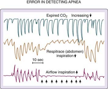
The vast majority of clinical sleep evaluations use qualitative measures of airflow for detecting SDB events. Extreme care must be taken when classifying respiratory activity because a patient’s airway can completely occlude during inspiration but release small puffs on expiration (detectable by thermistor or CO2 analyzer). Such events are erroneously categorized as hypopneas or even normal (unobstructed breathing). Figure 142-4 illustrates the problem. Airflow is recorded simultaneously with a CO2 analyzer and a pneumotachograph. During the obstructive apnea, periods of expiratory airflow occur (recorded by the pneumotachograph and the CO2 analyzer) in the absence of inspiratory flow (obvious in the pneumotachograph recording and unclear in the CO2 recording). Without the information from the pneumotachograph, the recording from the CO2 analyzer would be interpreted as evidence of uninterrupted inspiratory and expiratory airflow.


Methods to Detect Respiratory Effort


Qualitative airflow measures and devices monitoring expired gases cannot reliably differentiate between prolonged inspiration, central apnea, and obstructive apnea. Therefore, airflow is typically recorded in conjunction with measures of respiratory effort. The differential patterns of airflow and respiratory effort are commonly used to categorize apnea and hypopnea events as central, obstructive, or mixed.


Rib Cage and Abdominal Motion


Measuring rib cage and abdominal movement represents the most common technique for assessing respiratory effort in laboratory sleep studies. Rib cage and abdominal movement can be measured with a variety of techniques, including strain gauges, inductance plethysmography, and piezoelectric transducers. To determine whether respiratory effort is present, a single uncalibrated abdominal movement sensor will suffice.


Strain gauges are sealed elastic tubes filled with an electrical conductor through which an electric current is passed. When length is constant, current and resistance are constant. Stretching the strain gauge lengthens and narrows the cross-sectional area of the fixed-volume conductor. This produces a proportional increase in electrical resistance. Current varies inversely to the length of the gauge, thereby becoming an index of gauge length. A Whetstone bridge amplifier transduces this change to voltage so it can be displayed as a tracing showing rib cage or abdominal expansion (depending upon where it is placed).


Inductance pleythysmography electronically measures changes in the cross-sectional area of the rib cage and abdominal compartments by determining changes in inductance. Inductance is a property of electrical conductors characterized by the opposition to a change of current flow in the conductor. Transducers are placed around the rib cage and abdomen—the physiologic equivalent of conductors. Each transducer consists of an insulated wire sewn into the shape of a horizontally oriented sinusoid and onto an elasticized band.


Piezoelectric transducers are yet another method used to detect movement. These sensors can be placed on the rib cage and abdomen and are sensitive to changes in length.


During normal breathing, the major inspiratory muscles produce rib cage expansion and a downward movement of the diaphragm. These movements cause the pressure around and in the lung to become negative (relative to atmospheric pressure). The pressure gradient between ambient air and the lung draws air through the airways into the alveoli. Thus, a change in lung volume is the sum of the volume changes of the structures surrounding the lungs, the rib cage, and the abdomen.7 Other respiratory muscles (e.g., intercostal, sternocleidomastoid) also play a role in stabilizing the thoracic cage. Some clinicians erroneously interpret the abdominal and rib cage motion changes as implying separate activities of abdominal and thoracic respiratory muscles, but this is not the case. Virtually all the changes in abdominal and rib cage volumes (including paradoxical motion) can be explained by changes in the respiratory muscles directly inserting onto the thoracic cage. Paradoxical motion of the rib cage and abdomen can result from several changes, including loss of tone of the diaphragm, loss of tone of the other respiratory muscles, or upper airway obstruction (complete or partial). The mechanisms underlying this asynchronous motion of rib cage and abdomen are described in Box 142-2. Regardless of the pattern or its underlying mechanism, rib cage and abdominal movement reflect effort to breathe.


Box 142-2 Paradoxical Motion of the Rib Cage and Abdomen


Respiratory Muscle Electromyography


One of the older techniques for detecting respiratory effort entail recording intercostal muscle electromyographic (EMG) activity (Fig. 142-5). These uncalibrated recordings are made using standard surface electrodes placed in pairs in the intercostal space on the right anterior chest. Attaining an optimal signal requires practice, patience, and skill; recordings are prone to artifact, especially artifact from the electrocardiogram (ECG). Intercostal EMG recordings, when recorded properly, can be extremely valuable for differentiating central, obstructive, and mixed SBD events. Furthermore, although signals are not calibrated, cascading increases in respiratory effort are readily apparent from recordings.


Stay updated, free articles. Join our Telegram channel

Mar 13, 2017 | Posted by in NEUROLOGY | Comments Off on Monitoring Techniques for Evaluating Suspected Sleep-Disordered Breathing

Full access? Get Clinical Tree

Get Clinical Tree app for offline access