Multiple Sclerosis

46 Multiple Sclerosis


Clinical Vignette


A 23-year-old woman suddenly noted pain behind her right eye; this discomfort was exacerbated whenever she moved her eyes horizontally or vertically. Within 2 days, she lost central vision in her right eye; she could not read with that eye alone; however, her left eye was normal. She was otherwise perfectly healthy.


Neurologic examination demonstrated her visual acuity as 20/200 OD and 20/30 OS; associated findings confined to her right eye included a slightly irregular, poorly reactive pupil, diminished color vision, and papilledema. A magnetic resonance image (MRI) of the brain demonstrated a few periventricular demyelinating lesions. Treatment was begun with daily intravenous methylprednisolone infusions for 10 days. Her vision returned to normal within a few weeks.


She was well for another 4 years until the sudden onset of an annoying swollen and numb feeling in her left leg and concomitant occurrence of an electric shock–like sensation that spread down her back whenever she bent her neck. Examination demonstrated brisk muscle stretch reflexes, left greater than right, a left Babinski sign, ipsilateral decreased position sense, and contralateral diminished temperature appreciation from her right foot to her high thoracic region. MRIs demonstrated demyelination within her left cervical-thoracic spinal cord junction, and concomitant increased T2 signal within the corpus callosal and periventricular cerebral white matter. Although she once again responded well to methylprednisolone, and despite beginning ongoing therapy with beta-interferon, this young woman continued to have occasional exacerbations with some degree of remission. Eventually, she developed a persistent gait ataxia; nevertheless, she has maintained a very successful professional career, is married, and has a 3-year-old child.


Comment: This is a classic example of a previously healthy young person, whose initial optic neuritis held a relatively poorer prognosis as her brain MRI at that time showed signs of “silent” central nervous system (CNS) demyelination. Today, with that initial combination of optic neuritis and MRI findings, a diagnosis of multiple sclerosis (MS) would be made and an “ABC” medication begun at the outset of her illness.


Multiple sclerosis is the most common central nervous system disorder to affect young and middle-aged adults. Because the disease process has such protean manifestations and a variable course, demyelinating disorders have a broad clinical spectrum, ranging from a single benign episode to one that is potentially fatal. Epidemiologic studies demonstrate that MS is more prevalent in northern latitudes, twice as common in women, and most often presents during the third and fourth decades. Caucasians are twice as likely as people of color to acquire MS in the United States and Canada. Interestingly, when MS develops among Asian populations, it predominantly affects the optic nerves and spinal cord with much less involvement of the brain in contrast to North American and European persons. Individuals born in a northern latitude, who are race- and age-matched, gain the innate diminished risk of equatorial region habitats if they move south before age 15 years. Other persons who also move from the north to the south, but do not do so until after age 15 years, maintain their innate higher risks. Although there are some yet to be precisely identified genetic predisposing factors for MS, these alone cannot account for the above-defined variabilities, as genetically comparable populations vary in MS prevalence depending on place of birth and age of migration.


Pathology


With classic MS, the primary process is one of demyelination leading to loss of myelin from central nervous system (CNS) axons. Myelin loss (a nonspecific term) occurs concurrently with other pathologic processes that also affect the axons, glial elements, or vasculature. CNS oligodendrocytes are responsible for the elaboration of brain myelin. This is a predominantly lipid-based (70%) structure, with the remainder being protein based. One part, myelin basic protein, is particularly immunologically susceptible and experimentally encephalitogenic.


Inspection of the gross brain of an MS subject does not demonstrate any sign of abnormalities that would indicate the presence of the myriad histologic changes that are to be found on microscopic evaluation. However, the optic nerves, optic chiasm, and spinal cord may be atrophic to the native eye. Sometimes, areas of patchy demyelination are seen on the basis pontis surface, the cerebellar peduncles, and the surface of the medulla and floor of the fourth ventricle.


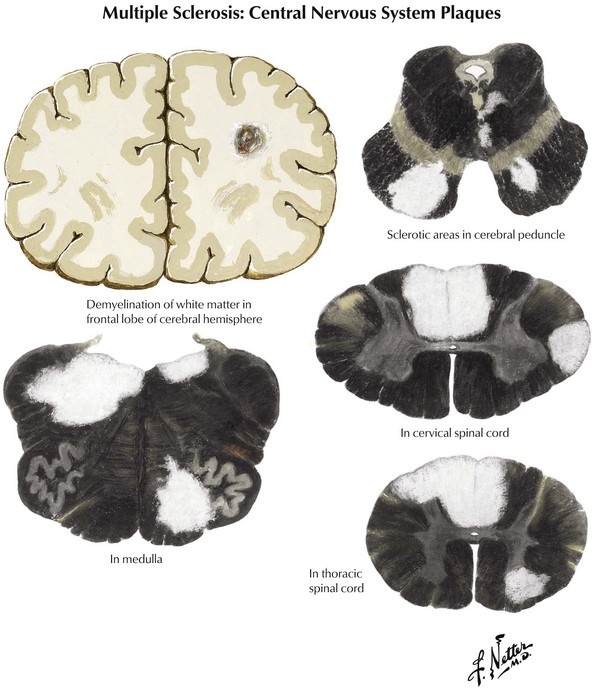
Coronal brain sections reveal changes similar to those noted on MRI, where variously sized MS plaques are apparent. Recently acquired lesions are pink and soft, whereas chronic MS lesions are gray, translucent, and firm (Fig. 46-1). It is often difficult to correlate the multiple lesions found at autopsy or by MRI throughout the neuraxis with a patient’s history. Sometimes classic MS plaques exist in patients who were never clinically suspected of harboring it.


Microscopic analysis demonstrates that many plaques have no relation to specific nerve tracts. Often, the plaques have a perivenular and paraventricular distribution. Myelin loss from a nerve fiber is distinct and best defined by toluidine blue stains. Macrophage accumulation is a frequent accompaniment. Active plaques contain myelin debris. Severe loss of oligodendrocytes within MS plaques is associated with the concomitant nonspecific finding of hypertrophic astrocytes. Signs of leptomeningeal inflammation, not unlike that found in acute disseminated MS, may be evident.


There is also a very significant component of axonal and neuronal damage in multiple sclerosis. This is particularly relevant to the long-term outcome and eventual disability. One can find evidence of axonal injury early on in the disease process. This is found in areas of obvious demyelination as well as areas of white and gray matter that appear normal to gross inspection. It is proposed that an antigen-specific destructive component related to both T cells and autoantibodies as well as the effects of activated macrophages and microglia lead to very significant axonal damage in the pathogenesis of MS. Mitochondrial function may also be impaired as various cellular substrates further contribute to this pathologic component.


Clinical Subtypes


The natural history of MS varies with the subtype of disease. Functional consequences may relate to some degree of axonal loss occurring after demyelination.


Benign MS


Clinical Vignette


A 52-year-old woman with migraine headaches since childhood, tobacco use throughout her adult years, and a history of transverse myelitis at age 14 years with complete recovery was subsequently noted to have a normal neurologic examination. She remained well until 65 years of age when she had an acute episode of left hemisensory deficit. An MRI scan of the brain revealed an acute lacunar infarct in the right thalamus on diffusion-weighted imaging, a chronic lacunar infarct in the left basal ganglia, and 10 bright lesions on T2 and FLAIR images, several of which were linear to ovoid in shape, configured perpendicularly to the ventricular walls, none enhancing with contrast. MRI of the cervical spine showed mild myelomalacia at C5 to C7 without other abnormality. She quit smoking and recovered with very little sensory impairment throughout her left side and lived without further neurologic symptoms until the age of 72 years, when she died of lung cancer.


Comment: This patient experienced a single bout of transverse myelitis as an adolescent, often a harbinger of more generalized MS. Medical technology at that time did not afford a simple way to explore her central nervous system for occult signs of multiple sclerosis. Clinical signs of more diffuse disease never developed. However, approximately 50 years later, when the patient required an MRI for a question of stroke, clinically inactive, more widespread MS became evident. In fact there was more than one explanation for the periventricular white matter abnormalities on MRI, namely, microvascular disease and migraine headaches. However, taken in the context of her prior episode of transverse myelitis, and the number and configuration of her white matter lesions, a diagnosis of relatively benign MS is likely. Certainly, whatever the idiosyncrasies of her immune system are that protected her, thus preventing further expression of more diffuse signs of clinical multiple sclerosis over her lifetime, once unlocked, they might provide the key for better appreciating the pathophysiology of MS and therefore direct more specific therapeutic research intervention in the future.


Relapsing–Remitting MS


Clinical Vignette


A 47-year-old woman sustained lower back and neck injury and concussion at 25 years of age in a motor vehicle accident. She underwent a C2-3 fusion and L4-5 diskectomy at that time and made a good recovery. She delivered two healthy children when she was ages 28 and 30 years. Five months after the birth of the first child, she began to experience numbness in the left leg and urinary urgency, with occasional leakage of urine. She was felt to have a recrudescence of her lumbar radicular symptoms and postpartum stress incontinence. Her primary care physician referred her for physical therapy, and her symptoms improved over the ensuing 2 months.


Just 1.5 years later, at age 32 years, she experienced increased difficulty lifting simple kitchen objects such as a milk carton and a frying pan and noted some numbness in her hands. She had increasing problems carrying her children, particularly placing them into their car seats. Her internist found Tinel signs at each wrist and thought she might have bilateral carpal tunnel syndrome; wrist splints were not helpful, and she remained relatively symptomatic. Over the years, she tried to remain active with her children, but increasingly found herself altering bicycle routes that they had once taken with ease as a family. She could not drink anything for 3 hours prior to going out on a bike ride to avoid urinary incontinence. Because she more commonly fatigued with any sustained walking, particularly during the warmer summer months, she curtailed her shopping.


Eventually she was referred to a neurologist after she developed a right foot drop, a subsequent series of falls, and then an exacerbation of her bladder symptoms. Examination demonstrated tandem ataxia, circumduction of her right leg, with diminished right arm swing, very brisk muscle stretch reflexes more on the right, sustained ankle clonus, bilateral Babinski signs, and markedly diminished vibration sense. Brain MRI revealed scattered bihemispheric periventricular and subcortical T2 and FLAIR hyperintensities without enhancement. MRI of the cervical and thoracic spine showed a stable C2-3 fusion, myelomalacia from T7 to T9 with patchy hyperintensity throughout this region, and an enhancing lesion at the conus. Cerebrospinal fluid examination demonstrated five oligoclonal bands without pleocytosis.


Comment: This is a relatively classic case of relapsing–remitting multiple sclerosis (RRMS) beginning subtly during the childbearing years. On occasions the onset may be more precipitous, almost mimicking a stroke; however, the clinical setting is with rare exceptions not quite as abrupt, and the findings do not replicate the damage one finds when a specific vessel is obstructed. Often, as in this instance, the symptoms are attributed to more benign entities such as various mononeuropathies, for example, the median nerve with her first visit to an internist, and to a peroneal lesion when the foot drop developed and she first visited an osteopath. Her limited exercise reserve, as exemplified by her diminished ability to walk or bike-ride any significant distance, could easily have been ascribed to fatigue, hysteria, or depression, something that commonly occurs during the first years of a demyelinating illness. This is particularly the case for a young woman who has recently taken on the new responsibility of caring for two babies. With time, the clinical setting clarifies itself.


Even the most skilled neurologist often had trouble confirming an MS diagnosis prior to the era of MRI. Today the brain MRI is sometimes normal or nonspecific early on, and it is not until one also obtains spinal imaging that the MS diagnosis is established. Lastly one needs to fastidiously maintain an objective approach to this type of patient, especially early on in the illness. It is important to be willing to reevaluate PRN and/or at a set interval. Corollary to this is that it is blatantly unfair to label the patient with new-onset symptoms as being histrionic when she has a normal neurologic examination and especially a normal brain MRI.


Secondary Progressive


Clinical Vignette


This now 50-year-old woman presented with a 29-year history of neurologic difficulties. She had reported having diplopia at age 21 years, making a good recovery. She was diagnosed with RRMS just 2 years later, after an episode of subacute right-sided weakness and cerebrospinal fluid (CSF) analysis demonstrating oligoclonal bands. She was treated with adrenocorticotropin hormone (ACTH). Although subsequently she required a right ankle–foot orthosis (AFO), she was able to return to an active life. In the early 1990s, she was prescribed subcutaneous interferon beta-1b that she tolerated poorly because of excessive injection-site reactions; she discontinued these within a few months. Subsequently she developed increasingly severe relapses every 1 to 2 years, each time undergoing treatment with intravenous (IV) methylprednisolone. She than began interferon beta-1a intramuscularly in the late 1990s when she visited a new neurologist because of increasing dependence on a cane and frequent bouts of urinary incontinence. Subsequently, mounting lower extremity weakness, spasticity, and fatigue evolved but she no longer experienced distinct flare-ups of her disease. It became increasingly difficult for her to keep up with the frequent travel that her work as an executive required.


Neurologic examination revealed an internuclear ophthalmoplegia (INO) and spastic paraparesis. Ultimately, the patient required an electric wheelchair to maintain mobility.


Comment: Typically, RRMS continues over many years, gradually leading to incremental decline in a range of neurologic functions resulting in varying degrees of neurologic disability. Eventually, the severity and discreteness of relapsing symptoms tend to wane, but dysfunction concomitantly becomes more insidious, resulting in secondary progressive MS (SSMS). Rarely, MS begins with a fulminant course followed by poor recovery, rapid progression, or both, ending in significant disability or death in a relatively short period after disease onset.


Primary Progressive


Clinical Vignette


A 45-year-old man who worked as a New York stockbroker had a gradual change in his commuting routine to and from work. For years, he would forego the subway for a brisk walk to the stock exchange after taking commuter rail from the suburbs. By the age of 40, he had universally taken to riding the train the entire distance. He attributed his “laziness” to aging and knee problems related to his high school basketball career. No longer could he keep himself out in front when bicycle-riding with his wife nor could he lift nearly the weight with his legs during his gym workouts that he had earlier on. To his embarrassment, he tripped on the stairs when walking with a coworker and knocked out his two front teeth.


Neurologic examination demonstrated a spastic gait with mild circumduction of the right leg, mild weakness of the iliopsoas bilaterally, brisk muscle stretch reflexes in the legs, bilateral Babinski signs, and loss of vibration perception at the toes, ankles, and knees.


Comment: In approximately 15% of patients with MS, the clinical course is more chronically progressive from the outset, in contrast to the more typical history of remissions and exacerbations of relapsing–remitting disease. This clinical setting defines another group of patients, those with primary progressive MS (PPMS). Most of these individuals have clinical evidence of significant spinal cord disease but a paucity of intracranial findings. Typically, progressive gait dysfunction and spasticity characterize their clinical course. Unlike RRMS, this subtype exhibits a male predominance, older age of onset, and poor response to disease-modifying strategies.


Therefore, the MS clinical spectrum can range from a relatively asymptomatic disorder, after an initial few benign episodes of CNS or optic nerve disorder, to an acute life-threatening illness that may mimic a brain tumor.


Differential Diagnosis


Because MS can affect any area of the CNS from the optic nerves to the distal spinal cord, patients present with extremely varied manifestations occurring over a wide age range from midchildhood into the early sixth decade. Whenever one is confronted with an acute neurologic process primarily affecting the CNS in a previously healthy young person, the physician must always consider if this could be the initial presentation of MS. There is a very broad differential diagnosis predicated on the patient’s initial symptomatology.


Ocular


Blurred vision, likely a manifestation of acute optic neuritis, is a very common initial symptoms of MS. This may have great variability in degree from gross total unilateral blindness to a subtle change in visual acuity. Vision changes may also be related to a poorly expressed diplopia secondary to an INO, one of the most common neuro-ophthalmologic manifestations of MS (Fig. 46-2). In the first instance, the differential diagnosis includes any process affecting the retina or cornea, and in the second, the diplopia may be related to a pseudo-INO, mimicking MS. This is typical of myasthenia gravis. Sometimes a true INO secondary to a stroke may occur in older hypertensive patients.


At times a subtle demyelinating lesion may be clinically uncovered by exposure to heat. For example, during a summer heat wave, or even just taking a shower, a patient may note relatively short-lived symptomatology, sometimes visual blurring or just a change in gait; this is known as Uhthoff syndrome. The symptoms are usually fleeting. These routinely disappear when the patient returns to more normal ambient temperatures.


The prognostic significance of an acute episode of optic neuritis (ON) has been the subject of numerous investigations. Today MRI provides an important means to put the ON event into a long term perspective. A recent ON study, with a median 6 years’ follow-up, demonstrated that slightly less than 50% of these patients had converted to clinically definite MS (CDMS). Most importantly, both the presence and the number of MRI-defined spinal cord lesions, and to a lesser extent gadolinium-enhancing lesions, as well as the number of infratentorial lesions at baseline were significant independent predictors of an even higher disability manifested as CDMS.


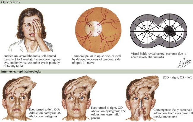
When temporally associated with a myelopathy, this clinical presentation is referred to as Devic syndrome, or neuromyelitis optica (NMO). This is a humorally mediated autoimmune demyelinating disorder that is immunologically distinct from MS. It is characterized by an autoantibody that recognizes aquaporin 4 (AQP4), a water channel expressed on astrocyte podocytes. NMO may sometimes have a poorer prognosis than MS although on occasion it is more acutely responsive to immunosuppression or chemotherapy. This Devic syndrome does not typically benefit from first-line long-term MS disease modifiers (Chapter 47).


Inner Ear or Cerebellum


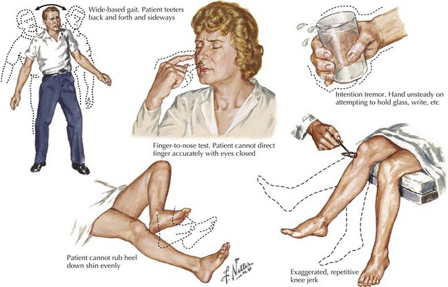
A vague feeling of unsteadiness, dizziness, and sometimes a whirling vertigo with nausea are frequent symptoms of MS. Precise definition of exactly what patients mean by “dizzy” is essential. Frequently, patients are in fact reporting a sense of dysequilibrium likely representing cerebellar dysfunction and less commonly reflecting vestibular pathways. Such patients typically demonstrate a broad-based gait, various signs of dysmetria, a tremor not seen in patients with a primary vestibular lesion, and often vertical nystagmus (Fig. 46-3).


The most common cause of true vertigo is either a primary vestibular neuronitis or the recurrent Ménière syndrome associated with hearing loss. These are both benign processes originating within the inner ear. However, one needs to always consider the possibility that the patient’s dizziness/vertigo has a primary CNS basis. One very important differential diagnostic clue to the latter is the finding of vertical nystagmus on the neurologic examination. This is not found when a peripheral mechanism for vertigo is present. The clinical demonstration of vertical nystagmus in a patient with vertigo requires an MRI to look for primary CNS disease.


Although spontaneous downbeat nystagmus is often the hallmark of another specific central nervous system lesion, namely an Arnold-Chiari malformation, this may also occur in MS patients reporting feelings of dizziness.


Myelopathies


Sensations of tingling, tightness, pins and needles, and electric shocks are common MS symptoms. Relatively early in the disease course, some of these patients report an electric shock–like sensation that usually radiates down the back or arms, occurring spontaneously when the patient bends his or her neck. This is known as Lhermitte’s sign (Fig. 46-4). This most commonly occurs with MS patients but often the patient does not report this per se. Rather the skilled examiner is trained to ask about such as it is pathognomonic and indicative of spinal posterior column dysfunction. Lhermitte’s sign can be reported with almost any form of spinal cord disease, including spondylosis with significant cervical spinal stenosis, a space-occupying lesion, vitamin B12 (cobalamin) deficiency, or copper deficiency syndromes, especially in individuals who have had major gastrectomies or unusual diets, such as excess zinc that blocks copper absorption.


Other patients with MS frequently report a tight bandlike distribution of numbness. When circumferential or hemicircumferential, particularly in the midtrunk, the immediate concern is a possible transverse myelitis or primary spinal cord mass lesion. Sometimes patients describe a distribution suggestive of a polyneuropathy with bilateral symmetric numbness. On other occasions, numbness may occur acutely in the hand, compatible with mononeuropathy or stroke. Other myelopathies deserving consideration relate to tropical spastic paraparesis or AIDS.


Clinical Vignette


A 58-year-old school teacher with a history of depression and sciatica noted the presence of left-sided weakness that progressed gradually over a year. She found it difficult to maneuver her left leg into a pair of pants and occasionally experienced buckling of the right leg when dressing. Her left-handed grasp of a pen was increasingly compromised and her handwriting was changing. Advancing fatigue was making it increasingly difficult for her to get through her work day. She began to experience difficulty driving due to stiffness and shaking of the right leg, and, eventually, she could not exit her car without lifting her right leg with her hands. This was associated with a band-like constriction sensation around her thorax and intermittent urinary incontinence that she attributed to child-bearing and age.


Neurologic examination demonstrated diminished fine coordinated movements of her dominant left hand, bilateral hand clumsiness, and right greater than left leg weakness, with increased tone distally in the lower extremities, and bilateral Babinski signs. Her gait was wide-based and she had a right foot drop. Sensory examination was normal except for mild symmetrically decreased vibration in the distal lower extremities.

Stay updated, free articles. Join our Telegram channel

Jun 4, 2016 | Posted by in NEUROLOGY | Comments Off on Multiple Sclerosis

Full access? Get Clinical Tree

Get Clinical Tree app for offline access