Fig. 44.1
Nasopharyngeal carcinoma (NPC). (a) Sagittal T1-weighted gadolinium-enhanced MR image. (b) Coronal T1-weighted gadolinium-enhanced image. A large enhancing mass is centered in the sphenoid sinus, replacing most of the clivus body. The pituitary gland is immediately above the mass
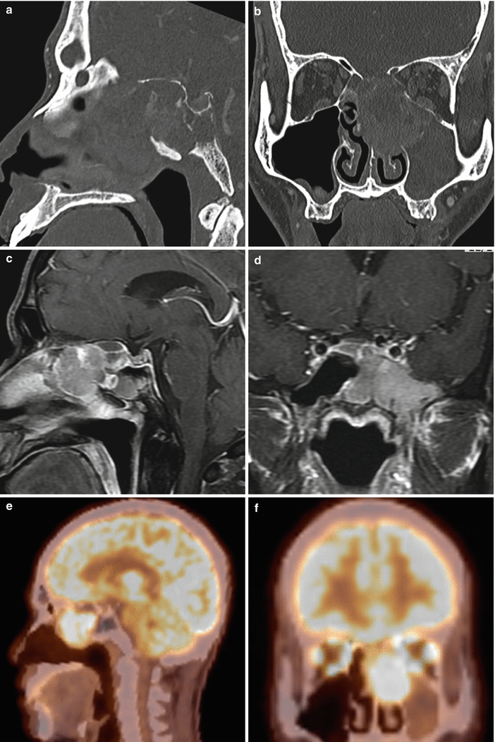
Fig. 44.2
NPC. (a) Sagittal CT image with contrast enhancement. (b) Coronal CT image with contrast enhancement. (c) Sagittal T1-weighted gadolinium-enhanced MR image. (d) Coronal T1-weighted gadolinium-enhanced image. A large enhancing mass is centered in the nasopharynx, eroding the septa and roofs of the ethmoid, the wall of the left maxillary antrum, and the upper body of the clivus. The mass extends laterally below the cavernous sinus to the floor of the middle cranial fossa and pterygopalatine fossa. The pituitary gland is distinctly seen above the mass. (e, f) FDG PET/CT shows the hypermetabolic sinus mass
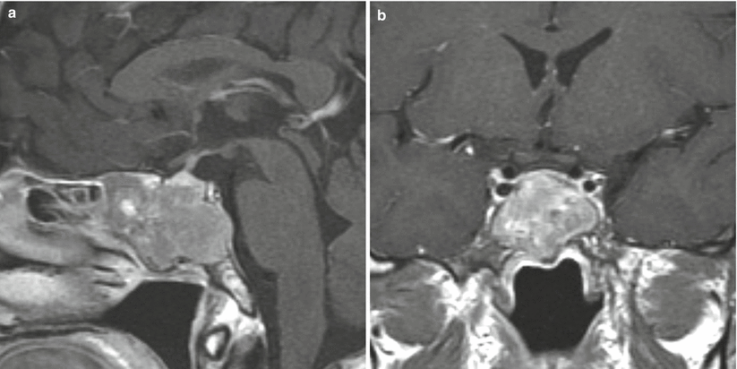
Fig. 44.3
Squamous cell carcinoma of the paranasal sinuses (PNSSCC). (a) Sagittal T1-weighted gadolinium-enhanced MR image. (b) Coronal T1-weighted gadolinium-enhanced image. An enhancing mass is centered in the sphenoid sinus, eroding the upper clivus, including the sellar floor. The pituitary gland is surrounded by the mass
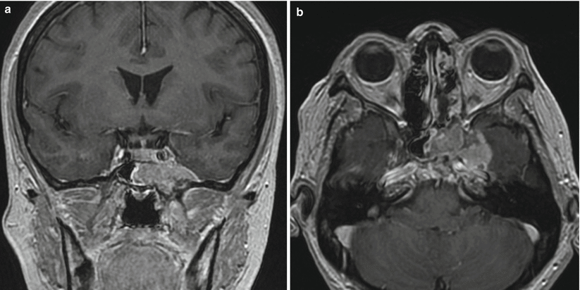
Fig. 44.4
PNSSCC. (a) Coronal T1-weighted gadolinium-enhanced MR image. (b) Axial T1-weighted gadolinium-enhanced image. An enhancing mass is seen centered in the left sphenoid sinus and middle fossa floor. The mass is highly invasive, with extension into the left medial/anterior temporal lobe
44.3 Histopathology
The current WHO classification of NPC includes keratinizing squamous cell carcinomas (WHO I) or nonkeratinizing carcinomas. Nonkeratinizing carcinomas are further subdivided into differentiated (WHO II) and undifferentiated (WHO III) carcinomas.
Latent EBV infection often leads to the development of NPC [1].
NPCs are characterized by polygonal epithelial cells intermixed with lymphoid cells.
PNSSCCs are often associated with (and may arise from) inverted epithelial papillomas of the paranasal sinuses [16].
They may be characterized by sheets of epithelial cells with varying degrees of keratin production, often with prominent palisading and intraepithelial nesting (Fig. 44.5) [8, 16].
Some PNSSCCs are associated with the human papillomavirus (HPV) [17].
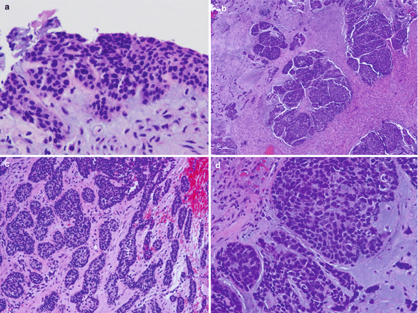
Fig. 44.5
Squamous cell carcinoma of the sphenoid sinus and sella. (a) Light microscopy revealed squamous dysplasia and carcinoma in situ of the mucosal surface (H&E, original magnification ×100). (b) Light microscopy revealed an invasive basaloid squamous cell carcinoma (BSCC). The BCSS grew as nested lobules in a fibrotic background. Large lobules showed compacted nests with a jigsaw-like pattern (H&E, original magnification ×40). (c) In this region, the BSCC grew as cords and trabeculae with peripheral palisading (H&E, original magnification ×100). (d) In a high-power view, the tumor was composed of closely packed, moderately pleomorphic basaloid cells with hyperchromatic nuclei and scant cytoplasm (H&E, original magnification ×200) (Adapted from Gu et al. [9], with permission)
44.4 Clinical and Surgical Management
In patients diagnosed with NPC, EBV DNA can often be detected by blood serology tests. The quantity of EBV DNA detected in blood indicates the stage and prognosis of the disease [1].
Standard TNM staging systems are typically applied at the time of diagnosis.
Endoscopic endonasal biopsy can often be performed to confirm the diagnosis.
Patients will commonly undergo biopsy or surgical debulking followed by radiation therapy and chemotherapy. Common chemotherapeutic regimens generally include cisplatin, 5-fluorouracil, and folinic acid [1, 20].
Intensity-modulated radiation therapy (IMRT) doses of greater than 66 Gy have been associated with improved progression-free survival [21].
The rate of hypopituitarism following radiation treatment for NPC is as high as 62 % in the 5 years following radiation. The growth hormone, cortisol, and thyroid hormonal axes were the ones most often affected. Vigilant monitoring and replacement of anterior pituitary hormones are recommended [1, 22].
The median overall survival for patients undergoing chemoradiotherapy was 80.3 months, versus 28.5 months with radiotherapy alone [3].
In a major series of patients with mostly stage III/IV NPC, the overall survival at 30 months was 97 % [20].Related posts:

Stay updated, free articles. Join our Telegram channel

Full access? Get Clinical Tree


