FIGURE 47.1 Lateral view of the cervical spine. Shows the vertebral bodies separated by intervertebral discs, the pedicles merging into the facet joint with its superior and inferior facets, and intervening pars interarticularis. The facets are oblique in the cervical region and more vertical in the lumbosacral spine. The uncovertebral joints are not true joints but just the opposing surfaces of the vertebral bodies. The uncovertebral processes may form osteophytes, or “spurs,” which then project into the foramen. (Used from Campbell WW. Essentials of Electrodiagnostic Medicine. Philadelphia: Lippincott Williams & Wilkins, 1999, with permission.)

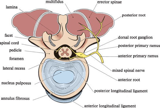
FIGURE 47.2 Cross section of a vertebral body with one pedicle cut away to show the contents of the intervertebral foramen, with the dorsal root ganglion lying in the midzone. Note the location of the multifidus muscle compartment and the innervation of the paraspinal muscles by the posterior primary ramus. The posterior longitudinal ligament is incomplete laterally, and disc ruptures tend to occur in a posterolateral direction. When a facet joint becomes enlarged due to osteoarthritis, it may encroach on the lateral recess, where the nerve root is entering the foramen. (Used from Campbell WW. Essentials of Electrodiagnostic Medicine. Philadelphia: Lippincott Williams & Wilkins, 1999, with permission.)
The mixed spinal nerve passes outward from the spinal canal through the intervertebral foramen. The foramen is a passageway formed by the vertebral body anteriorly, pedicles above and below and the facet mass and its articulation, the zygapophyseal joint, posteriorly. The neural foramen has an entrance, a middle zone, and an exit. The lateral recess of the spinal canal merges into the entry zone of the foramen. The dorsal root ganglion (DRG) occupies the midzone. The uncovertebral joints (of Luschka), which are not true joints, are the points where the posterolateral surface of a cervical vertebra comes into apposition with a neighboring vertebra. Degenerative osteophytes projecting into the intervertebral foramen from the uncovertebral “joints” may narrow it and cause radiculopathy (Figure 47.1). The uncovertebral joints are not present in the lumbosacral spine.
The tough anterior longitudinal ligament (ALL) extends lengthwise along the anterior aspect of the vertebral column, providing anterior reinforcement for the annulus. The posterior longitudinal ligament (PLL) extends along the posterior aspect of the vertebral bodies and reinforces the discs posteriorly. Compared with the ALL, the PLL is weak and flimsy and narrows as it descends. Disc herniations tend to occur posterolaterally, especially in the lumbosacral region, in part because of the lateral incompleteness of the PLL. In the cervical region, the PLL may ossify and contribute to spondylotic narrowing. The ligamentum flavum extends along the posterior aspect of the spinal canal. It buckles and folds during neck extension and may also contribute to canal narrowing.
The static anatomy of the spine provides only a partial understanding of the changes that occur on motion. Direct measurements have shown that the pressure within the disc varies markedly with different postures and activities. It is lowest when lying supine, increases by fourfold on standing, and increases a further 50% when leaning forward. The pressure is 40% higher sitting than standing. The higher pressure when sitting is clinically relevant, as patients with lumbosacral disc ruptures characteristically have more pain sitting than standing. The intradiscal pressure during a sit-up is astronomical.
The size of the intervertebral foramina decrease with extension and with ipsilateral bending. In extension, the facet joints draw closer together and the posterior quadrants of the spinal canal narrow. Cervical roots stretch with flexion and may angulate at the entrance to the foramen. The intraspinal subarachnoid pressure varies with respiration and increases markedly with Valsalva or restriction of venous outflow. The epidural and radicular veins change in size with posture and respiration. These dynamic changes, which are especially relevant in the presence of pathology, form the basis for clinical tests and historical questions useful for distinguishing the various causes for back and neck pain.
The Intervertebral Disc
The annulus provides circumferential reinforcement for the disc; the spherical NP allows the vertebral bodies above and below to glide and slip across it, like a ball bearing. The NP is eccentrically placed, closer to the posterior aspect of the disc (Figure 47.2). The relative thinness of the annulus posteriorly is another factor contributing to the tendency of disc herniations to occur in that direction. The great majority of the weight-bearing function of a normal disc is borne by the NP, which contains proteoglycans, macromolecules that heartily imbibe fluid. Early in life, the NP is 90% water, but it undergoes progressive desiccation over time. With desiccation of the nucleus and loss of compressibility, the annulus must assume more of the weight burden. This increased load, in the face of its own degenerative weakening, then makes the annulus prone to tears.
The Spinal Roots
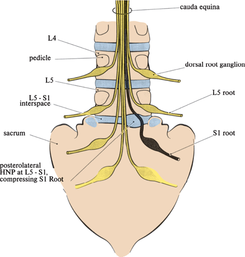
The anatomy of the spinal nerve is shown in Figure 24.3. Myotomal anatomy is discussed in Chapter 27 and dermatomal anatomy in Chapters 31 and 36. In the cervical spine, the nerve root exits over the vertebral body of like number until the C8 root exits beneath C7; all subsequent roots exit beneath the vertebral body of like number (Figure 24.1). In contrast to LSR, where disease usually affects the spinal root exiting one vertebral level lower, that is, disease at the L4-L5 level affects the L5 root, cervical radiculopathy (CR) tends to affect the nerve root laterally at its level of exit. Disease at the C5-C6 vertebral level affects the C6 root; at the C6-C7 level, the C7 root. When the cord terminates at the level of L1-L2, the remaining roots drop vertically downward in the cauda equina to their exit foramina. The L5 nerve root exiting at the L5-S1 interspace has arisen as a discrete structure at L1-L2 and had to traverse the interspaces at L2-L3, L3-L4, and L4-L5 before exiting at L5–S1, sliding laterally all the while. The L5 root could be injured by a central disc at L2-L3 or L3-L4, a posterolateral disc at L4-L5, or a far lateral disc or lateral recess stenosis at L5-S1 (Figure 47.3). A posterolateral disc at L4-L5 is the most likely culprit but not the sole suspect. The clinician must correlate the clinical localization of a given root lesion with the radiographic and clinical information to deduce the vertebral level involved and the proper course of action.

FIGURE 47.3 Posterior view of the cauda equina with exiting nerve roots. The nerve roots move laterally en route to their exit foramina. A posterolateral herniation of the nucleus pulposus (HNP) has compressed the S1 root as it passes by the L5–S1 interspace. A central HNP at any interspace could affect multiple roots. (Used from Campbell WW. Essentials of Electrodiagnostic Medicine. Philadelphia: Lippincott Williams & Wilkins, 1999, with permission.)
With aging and recurrent micro-and macrotrauma, degenerative spine disease develops. This involves both the disc (degenerative disc disease or DDD) and the bony structures and joints (degenerative joint disease or DJD). These processes are separate but related. Together, DDD and DJD are referred to as spondylosis. Small tears in the annulus may cause nonspecific, nonradiating neck or back pain. More extensive tears lead to disc bulging or protrusion, in which the disc herniates but remains beneath the PLL. Frank ruptures breach the PLL and allow a full-blown herniation of the nucleus pulposus (HNP) into the epidural space. Most HNPs occur in a posterolateral direction; occasionally, they are directly lateral or central. Which nerve roots are damaged depends largely on the direction of the herniation. In the face of disc herniation, the root may be damaged not only by direct compression but also by an inflammatory process induced by intradiscal proteoglycans, ischemia due to pressure, and adhesions and fibrosis.
The anterior elements, vertebral body and pedicles, normally bear 80% to 90% of the weight. As degenerative changes advance with desiccation and loss of disc height, the posterior elements (facets, pars, and laminae) may come to carry up to 50% of the weight-bearing function. This increases the work of the posterior elements and accelerates their degenerative changes. They react to the increased weight-bearing role by becoming hypertrophic and elaborating osteophytes. Osteoarthritis and synovitis of the facet joints is another point of pathology. In response to the increased loading attendant on loss of disc height and shift of weight bearing posteriorly, the facet joints develop degenerative changes: laxity of the capsule, instability, subluxation, and bony hypertrophy with osteophyte formation. The friction induced by minor instability and microtrauma leads to the formation of osteophytes. In the cervical spine, there is the added element of hypertrophy of the uncinate processes and the development of uncovertebral spurs. Degenerative osteophytes arising simultaneously from the uncus and from the vertebral body end-plate region may become confluent and create a spondylotic bar or ridge that stretches across the entire extent of the spinal canal. Like any arthritic joint, the facet may enlarge, impinging on the intervertebral foramen or the spinal canal, especially in the lateral recess. Loss of disc height causes the PLL and the ligamentum flavum to buckle and bulge into the canal. The degenerative changes in the discs and bony elements eventually produce cervical or lumbar spondylosis and may culminate in the syndrome of spinal stenosis.
All these degenerative changes leave less room for the neural elements. In the sagittal plane, the average cervical spinal cord is about 8 mm and the average cervical spinal canal about 14 mm. A sagittal canal diameter less than 10 mm may put the spinal cord at risk. The epidural space is normally occupied primarily by epidural fat and veins. When disc herniations and osteophytes intrude into the space, the resultant clinical manifestations depend in large part on how much room there is to accommodate them. Patients blessed by nature with capacious spinal canals can asymptomatically harbor a surprising amount of pathology. Patients with congenitally narrow canals and those who have undergone past spinal fusion procedures are at increased risk for developing spinal stenosis. Compression of vascular structures may introduce an additional complication of cord and/or root ischemia.
Several different clinical syndromes may ensue from degenerative spine disease, including the following: simple, single-level radiculopathy; multilevel radiculopathy; cauda equina syndrome; cervical myelopathy; cervical radiculomyelopathy; neurogenic claudication; lateral recess syndrome; and occasionally, a central cord or Brown-Sequard syndrome. Rarely, radiculopathy results from other processes, such as tumor (e.g., neurofibroma, meningioma, metastasis), arachnoid or synovial cysts, infection (e.g., Lyme disease, CMV, epidural abscess), infiltration (e.g., meningeal neoplasia, sarcoidosis), epidural block, irradiation, or ischemia (e.g., diabetes). A common cause of noncompressive radiculopathy is herpes zoster. Reactivation of a latent varicella-zoster virus resident in DRG cells triggers an H. zoster outbreak (“shingles”). Affected patients develop an extremely painful vesicular rash in the distribution of the involved dorsal root ganglia, usually a single dermatome. Although thoracic segments are involved most often, zoster can strike anywhere. Severe myotomal weakness may result when H. zoster strikes in the cervical or lumbar enlargements. Acute and chronic inflammatory demyelinating polyradiculoneuropathies produce marked abnormalities of the roots.
Because of the varied pathology involved, different types of radiculopathy occur in degenerative spine disease. The process is frequently multifactorial, involving some combination of disc herniation and spondylosis. The most straightforward clinical syndrome is unilateral “soft disc” rupture, an HNP. A similar clinical picture can result from a foraminal osteophyte, a “hard disc,” or spur. Some patients have soft disc superimposed on hard disc. It is clinically, radiologically, and sometimes surgically difficult to distinguish between soft disc and hard disc. Osteophytes, spurs, and foraminal stenosis are more common than simple soft disc in the etiology of CR. In Radhakrishnan et al.’s series, soft disc (i.e., not present in association with significant spondylosis) was responsible in only 22%; the remainder had hard disc or a combination. As a general rule, soft disc is more likely in younger patients. A central HNP may compress the spinal cord or cauda equina.
In compressive radiculopathy, sensory loss occurs in a dermatomal distribution and weakness in a myotomal distribution. Dermatomal sensory loss is less than expected because of extensive overlap in the innervation zones of spinal roots. Investigators used very different techniques to obtain the available dermatome maps (see Chapter 31 and Figure 36.5). The maps of Keegan and Garrett most closely approximate clinical reality. Sensory loss is most readily demonstrated in the signature zones of the major roots. Weakness in radiculopathy is also usually less than expected for a given myotome because most muscles receive multisegmental innervation (see Chapter 27). Disease affecting multiple levels causes much more severe weakness.
NECK AND ARM PAIN
Pain in the neck or upper extremity is a common clinical problem. Pain may involve the neck, shoulder, arm, forearm, or hand in virtually any combination. Potential causes of neck and arm pain are many. Common neurologic etiologies are CR, degenerative spine disease, brachial plexopathy, and peripheral nerve entrapment. Neck and arm pain of musculoskeletal origin is also common. Neurologic and musculoskeletal etiologies may be difficult to separate.
CERVICAL RADICULOPATHY
The population-based study of CR by Radhakrishnan et al. provided a wealth of interesting information. The incidence was highest at ages 50 to 54, with a mean age of 47, a male predominance, and a decline in incidence after age 60. There was a history of physical injury or exertion in only 15%; the most common precipitants were shoveling snow or playing golf. The onset was acute in half, subacute in a quarter, and insidious in a quarter, with the majority of patients symptomatic for about 2 weeks prior to diagnosis. Surgery was done in 26%. The disease tends to recur—some 31% of patients had a previous history of CR, and 32% had a recurrence during follow-up. At last follow-up, 90% of the patients had minimal to no symptoms. Others have noted this favorable long-term prognosis.
A number of clinical conditions may be confused with CR. These primarily include brachial plexopathies, entrapment neuropathies, and nonneuropathic mimickers. The more common musculoskeletal conditions causing confusion include shoulder pathology (bursitis, tendonitis, impingement syndrome), lateral epicondylitis, and DeQuervain’s tenosynovitis (see Chapter 48). Cervical myofascial pain, facet joint disease, and cervical vertebral body pathology can cause neck pain with referred pain to the arm. Pain can be referred to the neck, arm, or shoulder from the heart, lungs, esophagus, or upper abdomen.
Clinical Signs and Symptoms in Cervical Radiculopathy
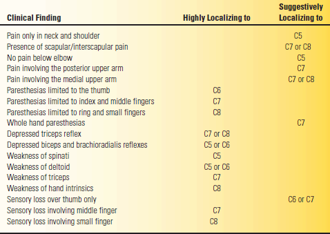
The classic articles by Yoss et al. and Murphey et al. detail the history and examination findings in CR. Yoss et al. evaluated 100 patients with surgically confirmed single-level cervical radiculopathies. The highly and suggestively localizing findings from these 100 patients are summarized in Table 47.1. Murphey et al. reviewed 648 cases of surgically treated singlelevel cervical radiculopathies. Findings in terms of pain radiation and neurologic deficits were similar to Yoss et al. Murphey et al. emphasized the occurrence of pain in the pectoral region in 20% of their cases; they opined that neck, periscapular, and pectoral region pain was referred from the disc itself and that arm pain was the result of nerve root compression.
TABLE 47.1 Clinical Findings in 100 Cervical Radiculopathy (CR) Patients

In the Radhakrishnan et al. series, cervicobrachial pain was present at the onset in 98% and was radicular in 65%. Paresthesias were reported by 90%, almost identical to the Yoss series. Pain on neck movement was present in 98%, paraspinal muscle spasm in 88%, decreased reflexes in 84% (triceps 50%, biceps or brachioradialis 34%), weakness in 65%, and sensory loss in 33%. In the Levin et al. series, 70% had motor and sensory symptoms, 12% had motor symptoms only, and 18% had sensory symptoms only.
The history, especially patterns of pain radiation and paresthesias, can provide localizing information in suspected CR. Radiating pain on coughing, sneezing, or straining at stool (Dejerine’s sign) is significant but seldom elicited. Increased pain on shoulder motion suggests nonradicular pathology. Relief of pain by resting the hand atop the head (hand on head sign) is reportedly characteristic of CR, but the author has seen this phenomenon with a Pancoast tumor. Hand paresthesias at night suggest carpal tunnel syndrome, but carpal tunnel syndrome can occur in association with CR (“double crush syndrome”), so nocturnal acroparesthesias do not exclude coexistent radiculopathy.
Physical examination in patients with suspected CR should include an assessment of the range of motion of the neck and arm, a search for root compression signs, detailed examination of strength and reflexes, a screening sensory examination, and probing for areas of muscle spasm or trigger points. Patients with either weakness or reduced reflexes on physical examination are up to five times more likely to have an abnormal electrodiagnostic study. A normal physical examination by no means excludes CR (negative predictive value 52%).
The cervical spine range of motion is highly informative. Patients should be asked to put chin to chest and to either shoulder, each ear to shoulder, and to hold the head in full extension; these maneuvers all affect the size of the intervertebral foramen. Pain produced by movements that narrow the foramen suggests CR. Pain on the symptomatic side on putting the ipsilateral ear to the shoulder suggests radiculopathy, but increased pain on leaning or turning away from the symptomatic side suggests a myofascial origin. Radiating pain or paresthesias with the head in extension and tilted slightly to the symptomatic side is highly suggestive of CR (Spurling’s sign or maneuver, foraminal compression test); brief breath holding or gentle Valsalva in this position will sometimes elicit the pain if positioning alone is not provocative. The addition of axial compression by pressing down on the crown of the head does not seem to add much. Spurling’s test is specific, but not very sensitive. Light digital compression of the jugular veins until the face is flushed and the patient is uncomfortable will sometimes elicit radicular symptoms: unilateral shoulder, arm, pectoral or scapular pain, or radiating paresthesias into the arm or hand (Viets’ sign). A slight cough while the face is suffused may increase the sensitivity. In the past, clinicians sometimes went so far as to put a blood pressure cuff around the patient’s neck to occlude the jugular veins (Naffziger’s sign). The two eponyms are often used interchangeably, and more often Naffziger’s sign is used for both techniques. Jugular compression is thought to engorge epidural veins or the cerebrospinal fluid reservoirs, which in the normal individual is harmless. But when some element of foraminal narrowing and nerve root pressure exists, the additional compression causes the acute development of symptoms. The same mechanism likely underlies the exacerbation of root pain by coughing, sneezing, and straining. Like Spurling’s test, Viets/Naffziger’s sign is specific but insensitive. It is less useful in lumbosacral than in CR.
An occasional CR patient has relief of pain with manual upward neck traction, particularly with the neck in slight flexion (cervical distraction test). Some patients have a decrease in pain with shoulder abduction (shoulder abduction relief test); this sign is more likely to be present with soft disc herniation. The mechanism is uncertain but probably related to the hand on the head sign. Flexion of the neck may cause Lhermitte’s sign in patients with cervical spondylosis or large disc herniations. Pain or limitation of motion of any upper-extremity joint should signal the possibility of nonradicular pathology. The differentiation of CR from primary shoulder disease (e.g., bursitis, capsulitis, tendonitis, or impingement syndrome) can be particularly difficult (see Chapter 48).
A focused but detailed strength exam should at least assess the power in the deltoids, spinati, biceps, triceps, pronators, wrist extensors, finger extensors, thenar muscles, and interossei. Testing muscles in a position of mechanical disadvantage may help detect mild weakness (see Chapter 27). The sensory exam should concentrate on the hand, and particularly assess touch, since the large, myelinated fibers conveying light touch are more vulnerable to pressure injury than the smaller fibers carrying pain and temperature. Reflex exam should include not only the standard upper-extremity reflexes but the knee and ankle jerks and plantar reflexes as well. Increased lower-extremity reflexes and extensor plantar responses suggest myelopathy complicating the radiculopathy.
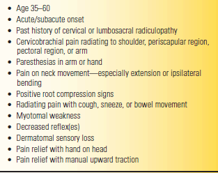
Based on the foregoing, Table 47.2 outlines the clinical data that favor the diagnosis of CR.
TABLE 47.2 Clinical Signs and Symptoms That Favor a Diagnosis of CR

Stay updated, free articles. Join our Telegram channel

Full access? Get Clinical Tree


