Neonatal Seizures


Neonatal Seizures


Robert R. Clancy

Eli M. Mizrahi


Neonatal seizures are a classic and ominous neurologic sign that can arise in any newborn infant. Their significance lies in their high incidence, association with acute neonatal encephalopathies, substantial mortality and neurologic morbidity, and the concern that seizures per se could extend the acute brain injury. Seizures in the neonate differ clinically and electrographically from those in mature infants and children. Diagnostic and treatment decisions remain limited by a paucity of rigorous scientific data for this population. This chapter reviews the significance of neonatal seizures, the pathophysiologic basis of clinical, electroclinical, and electrographic seizures, prognostic expectations, and etiologies, and surveys current treatment options that might themselves pose a risk to the developing brain.


HISTORICAL BACKGROUND

The appearance of “seizures,” “fits,” or “convulsions” in newborn infants has been known since antiquity. Seizure is derived from a Greek word implying a sudden “attack of disease.” The invention of the electroencephalograph by Hans Berger allowed investigators to discover the epileptic mechanisms that underlie seizure expressions in mature individuals. It was naturally assumed that clinical seizures in neonates were always associated with abnormal, excessive, paroxysmal electrical discharges arising from repetitive neuronal firing in the cerebral cortex. Despite the identification of electroclinical correlations of seizures in mature individuals, progress in understanding the nosology of neonatal seizures was only recently notable. Although some neonatal seizures were accompanied by simultaneous epileptic discharges demonstrated by electroencephalography, not every clinical event in a neonate that appears as an abrupt “attack” is truly epileptic, and the relationship between neonatal seizures and the conventional connotations of the term epilepsy demands careful scrutiny. Thus, seizures in the neonate are now distributed into three classes (Fig. 32.1). “Electroclinical” seizures are abnormal, clinically observable events that are consistently founded on a specific epileptic mechanism and coincide with an obvious electrographic seizure during simultaneous electroencephalographic (EEG) monitoring. “Clinical-only” seizures refer to other abnormal-appearing abrupt clinical events that are not associated with simultaneous electrographic seizure activity during EEG monitoring; they may be considered a type of nonepileptic seizure; “EEG-only” seizures lack definite clinical seizure activity; they are also called “subclinical” or “occult.”


SIGNIFICANCE OF NEONATAL SEIZURES


Incidence

The incidence of seizures in the first 28 days of life, one of the highest risk periods for seizures in humans, ranges between 1% and 5%. Depending on the methodology used, seizures occur at a rate of 1.5 to 5.5 per 1000 neonates (1, 2, 3, 4, 5, 6, 7), most within the first week of life (4). Incidence varies with specific risk factors. Lanska and colleagues (4) reported the incidence of seizures in all neonates to be 3.5 per 1000, but 57.5 per 1000 in very-low-birth-weight (<1500 g) infants, 4.4 per 1000 in low-birth-weight (1500-2499 g) infants, and 2.8 per 1000 in normal-birthweight (2500-3999 g) infants. Scher and colleagues (8,9) described seizures in 3.9% of neonates younger than 30 weeks conceptional age and in 1.5% of those older than 30 conceptional weeks.

The human newborn is especially vulnerable to a wide range of toxic or metabolic conditions. Sepsis, meningitis, hypoxic-ischemic encephalopathy (HIE), hypoglycemia, and hyperbilirubinemia are capable of eliciting seizures. This may explain, in part, the frequent occurrence of braindamaging events in the first 30 days of life. However, the neonatal brain itself may be especially prone to seizures when injured.


Figure 32.1 Three types of “seizures” in the newborn: “electrographic only,” “electroclinical,” and “clinical only.”

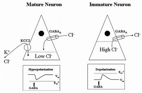
One suspected mechanism of enhanced seizure susceptibility in the newborn is the relative imbalance between inhibition and excitation. Compared with more mature brains, the neonatal brain has delayed maturation of inhibitory circuits and precocious maturation of excitatory circuits (10). This “imbalance” reflects a desirable and natural aspect of early central nervous system (CNS) development characterized by exuberant growth of excitatory synapses (10) coupled with activity-dependent pruning necessary for the prodigious rate of novel learning that faces all neonates. Moreover, according to studies in the neonatal rat, γ-aminobutyric acid (GABA) (the chief inhibitory neurotransmitter of the mature brain) may exert paradoxically excitatory effects in early CNS development (11,12). A developmentally dependent potassium chloride channel (KCC2) does not reach mature proportions in the rat hippocampus until the 15th postnatal day (Fig. 32.2). This age-dependent channel pumps potassium chloride from the interior of the neuron, reducing intracellular chloride levels. Thus, when the ligand-dependent GABA receptor is opened, extracellular chloride follows its electrochemical gradient into the neuron and hyperpolarizes it as expected. However, before the appearance of KCC2, the intracellular concentration of chloride is high, exceeding that of the extracellular space. In the immature rat, activation of the GABA receptor allows chloride to run along its electrochemical gradient out of the neuron, paradoxically depolarizing it (13).

Although neonatal seizures most commonly result from an underlying acute illness, some are reversible, the outward sign of a treatable condition. For example, the presence of hypocalcemia, hypomagnesemia, hypoglycemia, pyridoxine deficiency, or sepsis-meningitis may be heralded by neonatal seizures.


Prognostic Significance

Neonatal seizures are a powerful prognostic indicator of mortality and neurologic morbidity. The summary report from Bergman and associates (2) of 1667 patients noted an overall mortality of 24.7% before 1969 and 18% after 1970. Volpe (14) cited a mortality rate of 40% before 1969 and 20% after 1969. According to Lombroso (15), mortality decreased modestly from about 20% previously to 16% in the early 1980s. These improvements probably reflect better obstetrical management and modern neonatal intensive care. All of these studies relied on seizure diagnosis by clinical criteria and did not require EEG confirmation.


Figure 32.2 The effects of γ-aminobutyric acid may be paradoxically excitatory in early central nervous system development.

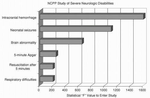
Survivors of neonatal seizures face an exceptionally high risk for cerebral palsy, often with mental retardation and chronic postnatal epilepsy. The National Collaborative Perinatal Population (NCPP) study (16,17) examined numerous clinical perinatal factors for their association with severe mental retardation, cerebral palsy, and microcephaly (Fig. 32.3). The clinical diagnosis of “neonatal seizures” was independently and significantly associated with these adverse outcomes and eclipsed only by “intracranial hemorrhage” in forecasting them. Neurologic functioning may even be impaired in those who appear “normal” after neonatal seizures (18).

Contemporary studies of the prognosis after neonatal seizures have emphasized the inclusion of infants whose seizure type was confirmed by EEG monitoring. Outcome has been assessed in terms of survival, neurologic disability, developmental delay, and postnatal epilepsy. Ortibus and colleagues (19) reported that 28% died; 22% of survivors were neurologically normal at an average of 17 months of age; 14% had mild abnormalities; and 36% were severely abnormal. Six years later, Brunquell and colleagues (20) put the mortality rate at 30%. Neurologic examinations showed abnormalities in 59% of survivors; 40% were mentally retarded; 43% had cerebral palsy; and 21% had postnatal epilepsy when followed up for a mean of 3.5 years.

Preliminary results of the Neonatal Seizures Clinical Research Centers from 1992 to 1997 have been reported (21). Of the 207 full-term infants with video-electroencephalography-confirmed
seizures who were prospectively enrolled, 28% died. Two-year follow-up data were available for 122 patients, or 86% of the survivors. Abnormal neurologic findings were noted in 42%. A Mental Developmental Index (MDI) score below 80 was present in 55%, a Psychomotor Developmental Index (PDI) score less than 80 in 50%, and chronic postnatal epilepsy in 26%.


Figure 32.3 The National Collaborative Perinatal Population (NCPP) study prospectively followed more than 34,000 mothers to identify perinatal events associated with adverse outcomes. Fifty neonates were found with subsequent severe neurologic handicaps. Six independent variables, including neonatal seizures, were associated with such neurologically devastating outcomes. (Adapted from Nelson KB, Broman SH. Perinatal risk factors in children with serious motor and mental handicaps. Ann Neurol 1977; 2:371-377, with permission.)

Whether seizures themselves adversely affect the developing brain is difficult to determine from clinical studies. Seizure burden may appear to influence outcome because some infants who experience brief, infrequent seizures may have relatively good long-term outcomes, whereas those with prolonged seizures often do not fare as well. However, easily controlled or self-limited seizures may be the result of transient, successfully treated, or benign CNS disorders of neonates, while medically refractory neonatal seizures may stem from more sustained, less treatable, or more severe brain disorders. Legido and associates (22) studied 40 neonates with electrographic seizures detected on randomly timed routine electroencephalogram examinations, and monitored them for cerebral palsy, mental retardation, and epilepsy. Overall neurologic outcome was more favorable in those with two or fewer seizures per hour than in those with more than that number. In the subgroup with seizures caused by asphyxia, cerebral palsy was more frequent when more than five seizures occurred per hour. However, these results might equally reflect more severe underlying injuries that triggered both the additional short-term seizures and greater morbidity on longterm follow-up. Attempting a balanced approach, McBride and coworkers (23) followed up 68 high-risk neonates with birth asphyxia, meningitis, and other stressors linked to neonatal seizures. All infants underwent long-term EEG monitoring. Forty developed electrographic seizures, and 28 did not. By logistic regression, electrographic neonatal seizures were significantly correlated with death and cerebral palsy. Other investigators (24), using proton magnetic resonance spectroscopy (1H-MRS) found an association of seizure severity with impaired cerebral metabolism measured by lactate/choline and compromised neuronal integrity measured by N-acetylaspartate/choline, and suggested this as evidence of brain injury not limited to structural damage detected by magnetic resonance imaging (MRI).


Neonatal Seizures May Be Inherently Harmful

Neonatal seizures may be intrinsically harmful to the brain (25). Most seizures were long assumed to be the innocuous, albeit conspicuous, result of an acute injury, and the subsequent long-term neurodevelopmental abnormalities, the result of their underlying causes, not the seizures themselves. Basic animal research into how extensively seizure activity may affect the developing brain has not resolved the controversy (26, 27, 28, 29, 30). Immature animals are more resistant than older animals to some seizure-induced injury (31). The immature brain may be resistant to acute seizure-induced cell loss (28); however, functional abnormalities such as impairment of visual-spatial memory and reduced seizure threshold (32) occur after seizures, and seizures induce changes in brain development, including altered neurogenesis (33), synaptogenesis, synaptic pruning, neuronal migration, and the sequential expression of genes including neurotransmitter receptors and transporters (34,35).


Figure 32.4 γ-Aminobutyric acid is a pentamer structure composed of six possible classes of subunits. The subunits themselves may have multiple variants that are expressed at different developmental ages.

Research by Holmes and colleagues (36) provides a newborn animal model for investigating whether recurrent seizures, induced by proconvulsant drugs that do not otherwise injure the brain, leave long-term undesirable effects on brain structure, learning, and susceptibility to spontaneous seizures. Starting on the first day of life, a series of about 25 seizures was induced by the administration of fluorothyl to neonatal rats. Behavioral testing in these animals as adults showed impaired spatial learning and memory, decreased activity levels, significantly lower threshold to pentylenetetrazol-induced seizures, and sprouting of CA3 mossy fibers, relative to controls that did not have neonatal seizures.

Neonatal seizures induce persistent changes in the inherent electrophysiologic properties of CA1 hippocampal cells in the rat (37). Although the resting membrane potentials in CA1 pyramidal neurons did not differ in controls and neonatal-seizure animals, reductions were noted in CA1 spike frequency adaptation and afterpolarization potentials after a spike train.


Figure 32.5 Rat pups subjected to seizures had significant differences in γ-aminobutyric acid subunit composition in later life compared with control animals. (Adapted from Brooks-Kayal AR, Shumate MD, Jim H, et al. Gamma-aminobutyric acid (A) receptor subunit expression predicts functional changes in hippocampal dentate granule cells during postnatal development. J Neurochem 2001;77:1266-1278, with permission.)

Seizures also accelerate neuronal death in the neonatal rat hippocampus in the setting of hypoxia (38). In the presence of hypoxia, the brain usually saves energy by blocking synaptic activity and electrically silencing the neuron; this effectively “turns off” the electroencephalogram. Seizures aggravate the hypoxic state by accelerating rapid anoxic depolarizations in the intact rat hippocampus. In effect, the generation of seizures “breaks the law of neuronal silence.”

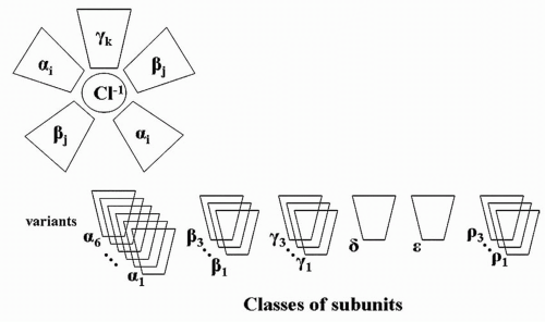
Finally, neonatal seizures in rats alter the subsequent composition of the GABAA receptor. GABAA is a pentamer in which five subunits assemble into a receptor structure (Fig. 32.4). Six subunit classes may comprise the pentamer: six variants of alpha, three of beta, three of gamma, one of delta, one of epsilon, and three of rho. The specific composition of an individual GABA
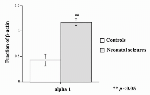
A receptor depends on developmental age. In the studies of Zhang and associates (12,39), rats with neonatal seizures had a substantially higher proportion of beta-actin in the α1 GABAA subunit component than did control animals (Fig. 32.5) (40).


CLASSIFICATION AND CLINICAL FEATURES OF NEONATAL SEIZURES

Application of a syndromic classification to neonatal seizures is limited when considered in light of the classification of the International League Against Epilepsy (ILAE) (41,42). Almost all neonatal seizures are thought to be symptomatic, an acute reaction or consequence of a specific
etiology. The ILAE addresses only five neonatal syndromes: benign neonatal convulsions, benign familial neonatal convulsions (BFNC), early myoclonic encephalopathy (EME), early infantile epileptic encephalopathy (EIEE), and migrating partial seizures of infancy. These are discussed later.

Seizures in the neonate are uniquely different from those in older infants and children. These differences are based on mechanisms of epileptogenesis and the developmental state of the immature brain and the relatively greater importance of nonepileptic mechanisms of seizure generation in this age group. Neonatal seizures may be classified by (a) clinical manifestations; (b) the relationship between clinical seizures and electrical activity on the electroencephalogram; and (c) seizure pathophysiology.


Clinical Classifications

A number of clinical classifications of neonatal seizures have been published (43, 44, 45, 46, 47, 48, 49). Early classifications focused on the clinical differences between seizures in neonates and those in older children: neonatal seizures were reported to be clonic or tonic, not tonic-clonic; when focal, they were either unifocal or multifocal. Later classifications included the term myoclonus. Another distinguishing feature of neonatal seizures is the occurrence of events described initially as “anarchic” (43) and thereafter “minimal” (45) or “subtle” (46). These events included oral-buccal-lingual movements such as sucking and chewing; movements of progression, such as bicycling of the legs and swimming movements of the arms; and random eye movements. First considered epileptic in origin, they were later deemed to be exaggerated reflex behaviors and thus were called “brainstem release phenomena” or “motor automatisms” (48). Table 32.1 lists the clinical characteristics of neonatal seizures according to a current classification scheme (50) that can be applied through observation.


Electroclinical Associations

Neonatal seizures may also be classified by the temporal relationship of clinical events to electrical seizure activity recorded on scalp electroencephalogram. In an electroclinical seizure, the clinical event overlaps with electrographic seizure activity. Some clinical-only events characterized as neonatal seizures may occur without any EEG seizure activity. Electrical-only seizures (also called subclinical or occult) occur in the absence of any clinical events.


Seizure Pathophysiology

Seizures may be classified as epileptic or nonepileptic (Table 32.2). Some clinical neonatal seizures are clearly epileptic, occurring in close association with EEG seizure activity, involving clinical events that can neither be provoked by stimulation nor suppressed by restraint, and directly triggered by hypersynchronous cortical neuronal discharges. The following properties of the developing brain intensify seizure initiation, maintenance, and propagation: increases in cellular and synaptic excitation and a tendency to enhance propagation of an epileptic discharge (29,31,51, 52, 53). The clinical events that are most clearly epileptic in origin are focal clonic, focal tonic, some types of myoclonic, and rarely spasms (Tables 32.1 and 32.2). Electrical-only seizures are, by definition, epileptic.

Best considered nonepileptic in origin (48,54) are events that occur in the absence of electrical seizure activity but that have clinical characteristics resembling reflex behaviors. Such clinical events, whether provoked by stimulation or arising spontaneously, can be suppressed or altered by restraining or repositioning the infant. The clinical events may grow in intensity with increases in the repetition rate of stimulation (temporal summation) or the sites of simultaneous stimulation (spatial summation). Some types of myoclonic events, generalized tonic posturing, and motor automatisms can be classified as “nonepileptic” (Tables 32.1 and 32.2).

Paroxysmal clinical changes related to the autonomic nervous system have been proposed as manifestations of seizures. These include stereotyped, episodic alterations in heart rate, respiration, and blood pressure (47,55,56). Skin flushing, salivation, apnea (57,58), and pupillary dilation may also be autonomic signs of seizures, but they are usually associated with other clinical manifestations, except in the therapeutically paralyzed infant (48).


Electrographic Seizures

Although clinical observation is critical to the detection of neonatal seizures, the electroencephalogram offers the most important means of confirmation and characterization. Infants with normal background activity are much less likely to develop seizures than are those with significant background abnormalities (59).


Interictal Background and Prediction Value

The ongoing cerebral electrical activity is the stage on which the drama of the episodic electrographic seizure unfolds. In many ways, the integrity of the EEG background is more critical than the mere presence or absence of the seizures themselves. For example, with or without electrographic seizures, an extremely abnormal EEG background (burst suppression [60] or isoelectric recording) inherently conveys a sense of profound electrophysiologic disruption and forecasts an exceedingly high risk for death or adverse neurologic outcome. Conversely, a nearly normal interictal EEG background suggests relatively preserved neurologic health despite the intrusion of the seizures.

The interictal background also occasionally can offer clues to seizure etiology. Persistently focal sharp waves may
suggest a restricted injury such as localized subarachnoid hemorrhage, contusion, or stroke, whereas multifocal sharp waves suggest diffuse dysfunction. Hypocalcemia is a consideration if a well-maintained background features excessive bilateral rolandic spikes. Inborn errors of metabolism, such as maple-syrup urine disease, are sometimes associated with distinctive vertex wicket spikes. Pseudoperiodic discharges raise the suspicion of herpes simplex virus encephalitis. A grossly abnormal electroencephalogram in the absence of any obviously acquired disease suggests cerebral dysgenesis.


TABLE 32.1 CLINICAL CHARACTERISTICS, CLASSIFICATION, AND PRESUMED PATHOPHYSIOLOGY OF NEONATAL SEIZURES


Classification


Characteristics


Pathophysiologic Basis


Focal clonic


Repetitive, rhythmic contraction of muscle groups of the limbs, face, or trunk
May be unifocal or multifocal
May occur synchronously or asynchronously in muscle groups on one side of the body
May occur simultaneously but asynchronously on both sides
Cannot be suppressed by restraint


Epileptic


Focal tonic


Sustained posturing of single limbs
Sustained asymmetric posturing of the trunk
Sustained eye deviation
Cannot be provoked by stimulation or suppressed by restraint


Epileptic


Generalized tonic


Sustained symmetric posturing of limbs, trunk, and neck
May be flexor, extensor, or mixed extensor/flexor
May be provoked or intensified by stimulation
May be suppressed by restraint or repositioning


Presumed nonepileptic


Myoclonic


Random, single, rapid contractions of muscle groups of the limbs, face, or trunk
Typically not repetitive or may recur at a slow rate
May be generalized, focal, or fragmentary
May be provoked by stimulation


Epileptic or nonepileptic


Spasms


May be flexor, extensor, or mixed extensor/flexor
May occur in clusters
Cannot be provoked by stimulation or suppressed by restraint


Epileptic


Motor Automatisms Ocular signs


Random, roving eye movements or nystagmus (distinct from tonic eye deviation)
May be provoked or intensified by tactile stimulation


Nonepileptic


Oral-buccal-lingual movements


Sucking, chewing, tongue protrusions
May be provoked or intensified by stimulation


Nonepileptic


Progression movements


Rowing or swimming movements
Pedaling or bicycling movements of the legs
May be provoked or intensified by stimulation
May be suppressed by restraint or repositioning


Nonepileptic


Complex purposeless movements


Sudden arousal with transient increased random activity of limbs
May be provoked or intensified by stimulation


Nonepileptic


Interictal EEG spikes per se have uncertain diagnostic significance (61). Interictal focal sharp waves and spikes are not typically considered indicators of epileptogenesis in the same way as they are in older children and adults. Compared with those of age-matched neonates without seizures (62,63), the interictal records of infants with electroencephalogram-confirmed seizures have background abnormalities, excessive numbers of “spikes” (lasting <200 milliseconds) compared with sharp waves (lasting >200 milliseconds), excessive occurrence of spikes or sharp waves per minute, and a tendency for “runs,” “bursts,” or “trains” of repetitive sharp waves. However, only a few infants with confirmed seizures exhibit all of these interictal characteristics, and many show no excessive spikes or sharp waves.


TABLE 32.2 CLASSIFICATION OF NEONATAL SEIZURES BY ELECTROCLINICAL FINDINGS


Clinical seizures with a consistent electrocortical signature (epileptic)


Focal clonic


Unifocal


Multifocal


Hemiconvulsive


Axial


Focal tonic


Asymmetric truncal posturing


Limb posturing


Sustained eye deviation


Myoclonic


Generalized


Focal


Spasms


Flexor


Extensor


Mixed extensor/flexor


Clinical seizures without a consistent electrocortical signature (presumed nonepileptic)


Myoclonic


Generalized


Focal


Fragmentary


Generalized tonic


Flexor


Extensor


Mixed extensor/flexor


Motor automatisms


Oral-buccal-lingual movements


Ocular signs


Progression movements


Complex purposeless movements


Electrical seizures without clinical seizure activity


Characteristics

At the heart of the epileptic process is the abnormal, excessive, repetitive electrical firing of neurons. Affected neurons lose their autonomy and are engulfed by the synchronized bursts of repeated electrical discharges. Sustained trains of action potentials arise in the affected neurons, which repeatedly fire and eventually propagate beyond their site of origin. At the conclusion of the ictus, inhibitory influences terminate the electrophysiologic cascade and end the seizure. Electrographic seizures in the neonate have varied appearances and are relatively rare before 34 to 35 weeks conceptional age. The morphology, spatial distribution, and temporal behavior of the seizure discharges may differ within and between individuals.


Morphology

An electrographic seizure is a discrete abnormal event lasting at least 10 seconds, with a definite beginning, middle, and end (64). No single morphologic pattern characterizes a seizure (Fig. 32.6). Even in the same patient, the ictal EEG activity may appear pleomorphic. The “typical” neonatal seizure begins as low-amplitude, rhythmic, or sinusoidal waveforms or spike or sharp waves. As the seizure evolves, the amplitude of the ictal activity increases, while its frequency slows. Spikes or sharp waves are not necessarily present. Instead, rhythmic activity of any frequency (delta, theta, alpha, or beta) can make up the ictal patterns at the scalp surface.


Spatial Distribution

In older children generalized seizures may appear simultaneously, synchronously, and symmetrically in both hemispheres. In the neonatal brain, which lacks the physiologic organization necessary for such exquisite orchestration, individual seizures always arise focally, for example, first appearing in the left temporal region (T3), migrating to adjacent electrode sites FP3, C3, or O1, and finally engaging the entire hemisphere (so-called hemiconvulsive seizure); seizures may also migrate from one hemisphere to another (50). Occasionally, simultaneous focal seizures may appear to behave independently, spreading to all brain regions, and superficially masquerading as a “generalized seizure.” However, the ictal patterns are not those of the truly generalized seizures, which usually are composed of spike or polyspike slow-wave discharges.

Although diffuse causes of encephalopathy such as meningitis, hypoglycemia, or hypoxia/ischemia may be expected to produce generalized seizures, each seizure instead arises from a restricted area of cortex. Multiple seizures that each originate from different scalp regions are called “multifocal-onset seizures”; those that arise from the same scalp location are unifocal onset and raise the possibility of a localized structural abnormality such as a stroke (65), reflecting the restricted functional disturbance.


Temporal Profile

An electrographic neonatal seizure lasts about 2 minutes and is followed by a variable interictal period (Fig. 32.7). These temporal characteristics were obtained from relatively brief tracings randomly selected during a variety of acute encephalopathies (9). Few studies comprehensively describe the natural history of electrographic seizures during continuous monitoring from the onset of acute neurologic illness. Solitary, prolonged electrographic seizures are rare in newborn infants; more than 90% of those seizures recorded in one study lasted less than 30 minutes (64). Repetitive brief serial seizures are much more characteristic than prolonged seizures lasting many hours.

Only gold members can continue reading. Log In or Register to continue

Stay updated, free articles. Join our Telegram channel

Oct 17, 2016 | Posted by in NEUROLOGY | Comments Off on Neonatal Seizures

Full access? Get Clinical Tree

Get Clinical Tree app for offline access