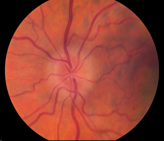
21 What are the basic components of the eight-point eye exam? 1. Best corrected visual acuity in each eye 2. Pupil exam 3. Ocular motility exam 4. Confrontation visual fields 5. Intraocular pressure 6. External exam 7. Slit-lamp exam 8. Funduscopic exam What is the appropriate way to check near visual acuity at the bedside with the near card? Check the vision at 14 inches from the patient. Measure and record the vision from each eye separately with patients wearing their glasses (including reading glasses if present). What is presbyopia, and how does it change how we test vision? Presbyopia is a decreasing ability to accommodate with age. In patients over the age of 40, it is important to provide some plus (e.g., +3.00 sphere) power lenses to help focusing up close.1–4 What does it mean if a patient’s vision is 20/20? ~20/80? In this notation, the numerator refers to the distance from the vision chart, and the denominator refers to the eye’s ability to see at that distance; 20/20 vision means that one can resolve at 20 feet what a normal person can resolve at 20 feet; 20/80 vision means that one can resolve at 20 feet what a normal person can resolve at 80 feet away. What are the basic components of an eyeglasses prescription? A typical prescription might read: OD: −1.50 +0.75 × 180 OS: −1.25 +0.50 × 150 OD means right eye (oculus dexter in Latin), and OS means left eye (oculus sinister in Latin). The first number indicates the spherical refractive error, meaning that it corrects for a single convergent or divergent refractive error in all meridians. The second number indicates any astigmatism, which means that parallel light rays are not converging on a single focal point on the retina, due to a difference in degree of refraction in different meridians. The astigmatic error is always listed at a specific axis, indicating the angle in degrees of the major astigmatic meridian, which is the third number in the prescription (e.g., in OD above, the +0.75 of astigmatic error is at axis 180 degrees). What are the major types of refractive error that a patient may have? 1. Myopia 2. Hyperopia 3. Astigmatism 4. Combination of spherical or astigmatic refractive errors What does it mean when a patient has myopia? Myopes typically have longer eyes (i.e., longer axial length) and are “nearsighted,” having a longer eye with a focal point in front of the retina. This is called axial myopia and the patient requires minus (−) lenses. There are also other nonaxial causes of myopia.1,3 What does it mean when a patient has hyperopia? Hyperopes have shorter eyes (i.e., shorter axial length) and are “farsighted,” having a smaller eye with a focal point behind the retina and would benefit from plus (+) lenses.1,3 What does it mean when a patient has astigmatism? This means that parallel light rays are not converging on a single focal point on the retina, due to a difference in degree of refraction in different meridians. The patient’s astigmatic error is typically listed at a specific axis (e.g., +3.00 at 180 degrees).1,3 What does it mean when a patient has anisometropia? This means the refractive error is different in each eye.1,3 What does the acronym PERRLA stand for? PERRLA is an acronym that can be used as a mnemonic device for pupils equal round and reactive to light and accommodation What can be misleading about the term PERRLA? PERRLA only tests the light and near response for the pupil and does not adequately comment on the presence or absence of a relative afferent pupillary defect (RAPD) or on the reaction of the pupil to dark (dilation under dark conditions). In a patient with a Horner syndrome, the anisocoria may only be detectable in the darker lighting condition as the smaller pupil fails to dilate as well. What is a relative afferent pupillary defect (RAPD)? An RAPD is a defect in the pupil pathway on the afferent side. The presence of an RAPD indicates a decreased pupillary response to light in one eye relative to the fellow eye. The RAPD is detected by swinging the light between the two eyes. In a patient with a right RAPD, the direct light reaction in the affected right eye will be less than the consensual response from the normal left eye and thus when the light swings from the unaffected eye back to the affected eye the affected right pupil will dilate, thus demonstrating the presence of the right RAPD.2–4 What does the Marcus-Gunn pupil refer to? The Marcus-Gunn pupil is another name for the RAPD. The presence of an RAPD is important to document as it suggests a problem in the ipsilateral retina or optic nerve in a patient with visual loss.2–4 What is anisocoria? Anisocoria means that the pupils are not of equal size. What is the most important step after anisocoria is detected? To determine if the anisocoria is worse in the light or the dark. Using PERRLA alone might miss the anisocoria in the dark. What is Horner syndrome? Horner syndrome refers to the clinical triad of ptosis, miosis, and anhidrosis due to a lesion in the ipsilateral oculosympathetic pathway. What is hippus? Hippus is rhythmic dilating and contracting movements of the iris that is a normal phenomenon and can be mistaken for an RAPD. What are ductions? Ductions are monocular rotations of the eye. • Adduction is movement of the eye nasally. • Abduction is movement of the eye temporally. • Elevation or supraduction is movement of the eye upward. • Depression or infraduction is movement of the eye downward. • Intorsion or incycloduction is nasal rotation of the superior portion of the vertical corneal meridian. • Extorsion or excycloduction is a temporal rotation of the superior portion of the vertical corneal meridian. What are versions? Versions are conjugate binocular movements. • Right gaze (dextroversion) is movement of both eyes to the right. • Left gaze (levoversion) is movement of both eyes to the left. • Upgaze (elevation) is movement of both eyes upward. • Downgaze (depression) is movement of both eyes downward. • Dextrocycloversion is right rotation of the superior portion of the vertical corneal meridian of both eyes. • Levocycloversion is left rotation of the superior portion of the vertical meridian of both eyes. Vergences, on the other hand, are disconjugate binocular movements. • Convergence is when both eyes move medially. • Divergence is when both eyes move laterally. What are the six extraocular muscles and their innervations? Lateral rectus: sixth nerve (abducens) Superior oblique: fourth nerve (trochlear) Medial rectus: third nerve (oculomotor) Superior rectus: third nerve (oculomotor) Inferior rectus: third nerve (oculomotor) Inferior oblique: third nerve (oculomotor) The eye is “down and out,” with ptosis, and a fixed and dilated pupil.3,4 One must rule out an aneurysm of the posterior communicating artery compressing the third cranial nerve! What is the most optimal test for evaluation of a new complete third nerve palsy? Computed tomography angiography (CTA) is a good first test for excluding an aneurysm but an MRI with and without contrast might still be needed for nonaneurysmal causes of third nerve palsy. What is the difference between a pupil-sparing and pupil-involving third nerve palsy? The parasympathetic fibers providing pupillary input travel externally on the nerve. The small vessels affected by microvascular disease travel internally within in the nerve. Pupil-involving third nerve palsy is secondary to a compressive condition such as an aneurysm until proven otherwise; it can be secondary to tumor, inflammation, infection, infiltration, or trauma. Pupil-sparing third nerve palsy refers to normal pupillary function but complete loss of lid and ocular motor functions of the third nerve. This is typical of an ischemic etiology (e.g., diabetes), with a common course of resolution in 3 months. Partial third nerve palsy refers to incomplete involvement of the eyelid and ocular motor function in a third nerve palsy, and can occur without involvement of the pupil, which is different from true pupil sparing in an otherwise complete third nerve palsy. Partial third nerve palsies should be evaluated for an underlying etiology. How do patients with a fourth nerve palsy present? Patients present with binocular vertical diplopia, typically worse in downgaze, and may have associated head tilt. Why is the fourth cranial nerve particularly susceptible to trauma and what are some other potential causes? The fourth cranial nerve has the longest intracranial course. It is important to remember that trauma, ischemia, and decompensation of a congenital fourth nerve palsy are common etiologies. Old photos might demonstrate a long-standing head tilt. Large vertical fusional amplitudes are also suggestive of a long-standing or congenital process. What is the diagnostic approach to a fourth nerve palsy? The Parks-Bielschowsky three-step test can help in the diagnosis of a fourth nerve palsy. • The first step is to determine which side is hypertropic (visual axis higher). • The second step is to determine in which gaze (left or right) the hypertropia is worse. • The third step is to determine which head tilt makes the hypertropia worse. A right fourth nerve palsy is a “marching palsy” (right, left, right)! The patient will have a right hypertropia, worse on left gaze, and worse on right head tilt. • A “fourth step” confirms that the torsion matches for the involved fourth nerve palsy (ispilateral excyclotorsion) rather than alternative etiologies (e.g., skew deviation).3,4 What is the classic presentation of a sixth nerve palsy? Horizontal diplopia that worsens on ipsilateral gaze, especially when viewing in the distance, often associated with an esodeviation (eye turned inward) in primary gaze that is worse on gaze toward the paretic lateral rectus muscle. What is important to rule out with new sixth nerve palsy? Lesions of the pons, subarachnoid space (nonlocalizing sign), cerebellopontine angle (e.g., acoustic neuroma or meningioma), cavernous sinus, superior orbital fissure, or orbit may present with a sixth nerve palsy. One should always evaluate if the sixth nerve palsy is isolated or nonisolated (e.g., checking for other neurological findings including CN V and CN VII in any new sixth nerve palsy). What are other important causes of sixth nerve palsies? Ischemia (e.g., diabetes), elevated intracranial pressure, compressive lesions anywhere along the course of the sixth nerve, inflammation (e.g., chronic inflammation of the petrous bone in Gradenigo syndrome), giant cell arteritis What is a gaze palsy? This is also known as conjugate ophthalmoplegia and refers to a symmetric limitation of movements of both eyes in the same direction. For example, a horizontal gaze palsy would indicate that the patient cannot look to the left or to the right. What causes a horizontal gaze palsy? This can be from a contralateral supranuclear lesion of the frontal eye fields in the cerebrum or an ipsilateral brainstem lesion involving the parapontine reticular formation (PPRF) or sixth nerve nucleus in the pons. Myogenic and neuromuscular junction (e.g., myasthenia gravis) defects can also produce gaze palsies. What is the role of the doll’s-eye maneuver? The doll’s-eye maneuver tests the integrity of the infranuclear pathways, similar to caloric stimulation. If the doll’s-eye maneuver overcomes a horizontal or vertical gaze palsy, then think supranuclear lesion. If it does not overcome the deviation, think brainstem nuclear or an infranuclear lesion. What is Parinaud’s syndrome? Parinaud’s syndrome is also known as dorsal mid-brain syndrome, characterized by paralysis of upgaze, light-near dissociation of pupils, convergence-retraction nystagmus (aka nystagmus retractorius), eyelid retraction (Collier’s sign), and conjugate down gaze in the primary position (setting-sun sign). Who is typically affected by Parinaud’s syndrome? Classically patients with pineal region tumors (e.g., germinomas, pineal tumors), although it can also be patients with multiple sclerosis, strokes, or other damage affecting the dorsal midbrain region. Hydrocephalus resulting in increased ICP will also produce downward gaze. What is internuclear ophthalmoplegia? Gaze disorder characterized by adduction deficit of one eye and dissociated nystagmus of the abducting eye in attempted horizontal gaze.3 What can cause internuclear ophthalmoplegia? A lesion of the medial longitudinal fasciculus ipsilateral to the adduction deficit, classically secondary to multiple sclerosis. What is myasthenia gravis (MG)? An immunological disorder involving antibodies to acetylcholine receptors (AChRs), leading to variable muscle weakness.3 What are clinical findings associated with myasthenia gravis? Ptosis Fatigability: worsens throughout the day; may be elicited by having the patient sustain upgaze for 1 minute. Variability: repeated evaluation may reveal different measurements. Enhancement of ptosis: elevating the ptotic eyelid causes the less ptotic eyelid to fall. Cogan’s eyelid twitch: A saccade from downgaze to upgaze elicits over-elevation of the upper eyelid. Diplopia due to the involvement of any one extraocular muscle or a combination of extra-ocular muscles. MG can mimic any pattern of ophthalmoplegia. MG does not involve the pupil, cause pain, or loss of sensory function. Systemic muscle involvement can produce generalized weakness and speaking or swallowing difficulty. What is the diagnostic workup for myasthenia gravis? Clinical evaluation can include: • Sleep test: improvement of ptosis after 30 minutes of rest suggests MG. • Ice-pack test: improvement of ptosis after 5 to 10 minutes of an ice pack applied to the eyelids suggests MG. • Tensilon test: improvement of ptosis 60 seconds after injection of 2 mg (0.2 cc) of edrophonium chloride, or after subsequent injection of up to two 4-mg doses suggests MG. • Chest CT scan for thymoma • Laboratory studies should include antibodies to acetylcholine receptor antibodies. Binding antibodies are ordered first. If these screening antibodies (Ab) tests are negative, additional blocking Ab, modulating Ab, or in some cases (especially generalized seronegative MG) anti-MuSK (muscle-specific kinase) Ab may be ordered. • Single-fiber electromyography is another sensitive test for MG. What is the direct ophthalmoscope and how is it used? • The direct ophthalmoscope is a handheld instru ment used to examine the optic disk as well as the posterior pole, giving approximately 15× magnification. • When using the direct ophthalmoscope, remember to align on the patient’s right side and use your right hand to examine the patient’s right eye, and align on the patient’s left side and use your left hand to examine the patient’s left eye. • The first step is to evaluate the red reflex from approximately 2 feet back. With the dial (Rekoss disk) set on 0, the examiner should approach the patient’s eye, with the thumb of the free hand raising the upper eyelid and the ulnar border of the hand holding the instrument against the patient’s cheek. • It may be necessary to dial the focusing lenses to clarify the fundus image, depending on the patient’s and examiner’s refractive error. • Once clear, the next step is to find the optic disk by following a retinal blood vessel. • The vascular bifurcations tend to form an arrow that points to the disk. • Then evaluate the peripapillary retina, follow the blood vessels outward from the disk, and finally the macula (retina between the vascular arcades). What is optic disk edema? What distinguishes optic disk edema from pseudopapilledema? • True optic disk edema is characterized by blurred disk margins that obscure the peripapillary nerve fiber layer and the retinal vessels as they cross the disk head. • Optic disk hyperemia may be present due to congestion of the disk microvasculature, and dilation and telangiectasia of the surface disk capillaries. • Obligatory pathological findings in true disk edema include retinal hemorrhages, exudates, cotton wool patches, or peripapillary subretinal fluid. • Pseudopapilledema does not produce edema of the peripapillary nerve fiber layer and is often due to underlying congenital calcified hyaline bodies called optic disk drusen. These do not obscure the peripapillary blood vessels and do not produce any of the obligatory signs. Fig. 21.1 Pseudopapilledema with slightly blurred disk margins but no retinal vessel obscuration or surface capillary dilation.
Neuro-Ophthalmology
21.1 Eight-Point Exam and Visual Acuity
21.2 Pupils
21.3 Ocular Motility
21.4 The Optic Disk
![]()
Stay updated, free articles. Join our Telegram channel


Full access? Get Clinical Tree


